-
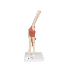 This high quality, life-size functional elbow joint model provides a graphic demonstration of the anatomy and mechanics of the human elbow joint. Use this fully flexible model to demonstrate abduction, anteversion, retroversion and internal/external rotation. Elbow joint consists of portion of the humerus, complete ulna and radius as well as joint ligaments. Comes on removable stand for easy study or display.
3B Scientific Functional Human Elbow Joint Model with Ligaments – 3B Smart Anatomy
-
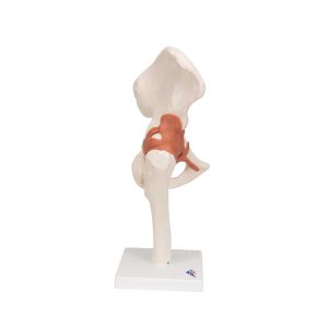 This life size functional hip joint model clearly shows the anatomy and mechanics of the human hip joint. This fully flexible hip joint demonstrates abduction, anteversion, retroversion and internal/external rotation. This high quality functional joint consists of a portion of femur, hip bone and joint ligaments. Comes on a stand for easy display in the classroom or doctor’s office.
3B Scientific Functional Human Hip Joint Model – 3B Smart Anatomy
-
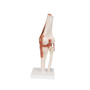 This high quality, life-size functional knee joint model clearly shows the anatomy and mechanics of the knee joint. This fully flexible knee joint model demonstrates abduction, anteversion, retroversion and internal/external rotation. The model consists of portion of femur, tibia and portion of fibula; also includes meniscus, patella with quadriceps tendon and joint ligaments, including the ACL and PCL. Delivered on removable stand for easy study or display.
3B Scientific Functional Human Knee Joint Model with Ligaments – 3B Smart Anatomy
-
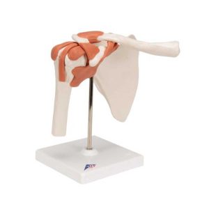 This high quality life-size functional shoulder joint model shows the anatomy and mechanics of the shoulder joint. Consisting of the scapula, clavical, portion of humerus and joint ligaments, this fully flexible shoulder joint model clearly demonstrates abduction, anteversion, retroversion and internal/external rotation. The Functional shoulder joint model comes on a stand for easy study and display.
3B Scientific Functional Human Shoulder Joint – 3B Smart Anatomy
-
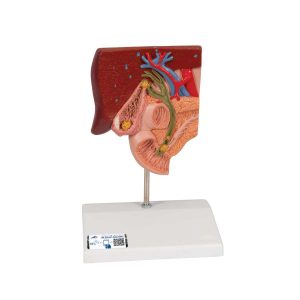 This graphic gallstone model for patient education shows the anatomy of the biliary system and its surroundings in half natural size. Both acute inflammation (cholecystitis) and the tissue changes caused by chronic inflammation can be identified in the gallbladder wall. Gallstones can be found in the following typical locations: In the fundus area of the gall bladder In the area of the spiral valve In the area of the common bile duct In the papillary opening to the small intestine Gallstone model mounted on base.
3B Scientific Gallstone Model – 3B Smart Anatomy
-
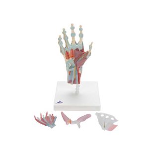 The bones, muscles, tendons, ligaments, nerves, arteries, and veins are all featured in this high quality 4 part model of the hand and lower forearm. The dorsal side of the hand shows the extensor muscles as well as portions of the tendons at the wrist as they pass under the extensor retunaculum. The palmar face of the hand is represented in three layers, the first two are removable to allow detailed study of the deeper anatomical layer of the hand. In addition clinically important structures such as the median nerve and superficial palmar arterial arch can be explored in detail in the hand model. The deepest anatomical layer allows for study of the intrinsic muscles and deep palmar arterial arch in addition to other details of the anatomy of the hand. This high quality anatomically correct hand model with ligaments and muscles is great for detailed study.
3B Scientific Hand Skeleton Model with Ligaments and Muscles – 3B Smart Anatomy
-
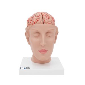 This C25 deluxe brain comes with opened head to allow detailed study of the brain‘s position in the skull. The human head is horizontally divided above the skull base. This medially divided deluxe brain model shows the brain arteries in detail. Both halves of this brain model can be disassembled into: Frontal with parietal lobes Temporal with occipital lobes Half of brain stem Half of cerebellum The classic brain is a great tool for education on the human nervous system and anatomy of the brain. The brain in a head base is delivered on a base.
3B Scientific Human Brain Model with Arteries on Base of Head, 8 part – 3B Smart Anatomy
-
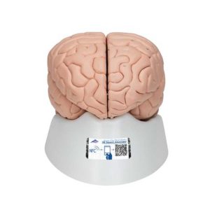 A very detailed model of the human brain which is medially divided. Both halves of this brain can be disassembled into: Frontal with parietal lobes Temporal with occipital lobes Half of brain stem Half of cerebellum This brain is a great educational tool for teaching and learning about the human nervous system and anatomy of the brain. The detailed brain is delivered on a removable base for easy display in the classroom.
3B Scientific Human Brain Model, 8 part – 3B Smart Anatomy
-
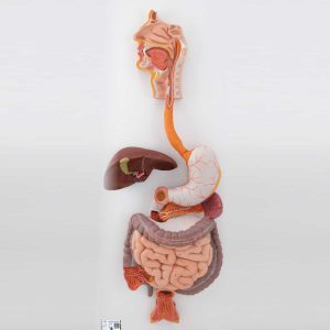 Life-size human digestive system model that demonstrates the entire digestive system in graphic relief. Digestive system features: Nose Mouth cavity and Pharynx Esophagus GI tract Liver with gall bladder Pancreas Spleen The duodenum, caecum and rectum of the digestive system are opened. The transverse colon and front stomach wall are removable from the digestive system for detailed study of the anatomy. This high quality digestive system is a great teaching tool for any anatomy lesson or doctor’s office. Digestive system mounted on baseboard for easy display in the classroom.
3B Scientific Human Digestive System Model, 3 part
-
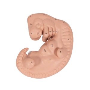 This human embryo model shows the anatomy of an embryo at approximately 4 weeks old. At 25 times life size this human embryo is great for studying human development. The high quality model is affordable without sacrificing any anatomical detail.
3B Scientific Human Embryo Model, 25 times Life-Size – 3B Smart Anatomy
-
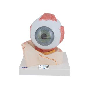 Removable parts of this anatomical human eye model include: Upper half of the sclera with cornea and eye muscle attachments Both halves of choroid with iris and retina Lens Vitreous humour This eye model is great for studying the anatomy of the human eye! Eye on base of bony orbit.
3B Scientific Human Eye Model, 5 times Full-Size, 7 part
-
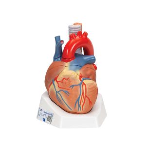 This heart model shows the anatomy of the heart model and is horizontally sectioned at the level of the valve plane. The following parts can be removed from the heart: Esophagus Trachea Superior vena cava Aorta Front heart wall Upper half of the heart This high quality model clearly shows over 30 different anatomical regions in the heart. Comes the product manual for easy identification of anatomical features.
3B Scientific Human Heart Model, 7 Part – 3B Smart Anatomy
-
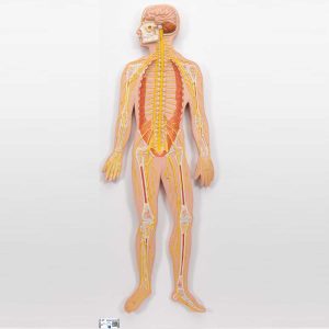 This nervous system relief model shows a schematic representation of the central and peripheral nervous system. An excellent nervous system model to study the structure of the human nervous system. This nervous system model is 1/2 life size and is a great teaching tool. The nervous system model is delivered on baseboard.
3B Scientific Human Nervous System Model, 1-2 Life-Size – 3B Smart Anatomy
-
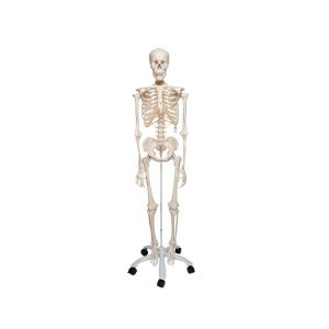 Proven Quality – even more stable! Stan, the standard model of a human skeleton, has been appreciated throughout the world for decades. Thanks to its very high quality and robust construction, it is perfect for use in hospitals, schools, universities and laboratories. The other advantages of the 3B Scientific® skeleton are: • Exceptional value for money • 3 year guarantee • Top quality natural casting • Final assembly carried out by hand • Made from a durable, unbreakable synthetic material • On a stable metal stand with 5 casters (painted white) • Close to the realistic weight of around 200 bones • Natural skeleton size • 3 part assembled skull • Individually inserted teeth • Limbs can be removed quickly and easily • Skull with magnetic connections Comes with metal stand and transparent dust cover.
3B Scientific Human Skeleton Model Stan – 3B Smart Anatomy
-
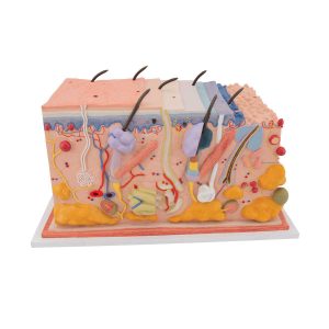 This unique skin model shows a section of human skin in three dimensional form. Individual skin layers are differentiated, and important structures of the skin such as hair, sebaceous and sweat glands, receptors, nerves and vessels are shown in detail. The high quality skin block model is mounted on baseboard. Demonstrating the anatomy of the human skin has never been easier! This skin block model details the human skin in 70 times life size.
3B Scientific Human Skin Section Model, 70 times Full-Size – 3B Smart Anatomy
-
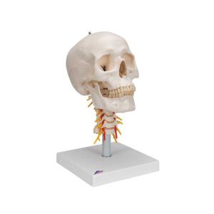 This human skull replica is flexibly mounted on a cervical spine. Features also shown are the hindbrain, spinal cord, cervical nerves, vertebral arteries, basilar artery and rear cerebral arteries. Skull can be removed from stand. High quality original human skull cast Skull is handmade from hard, unbreakable plastic Highly accurate representation of the fissures, foramina, processes, sutures etc. Can be disassembled into skull cap, base of skull and mandible Mandible of skull is mounted on a spring to easily demonstrate natural movement Add this detailed human skull model to your collection today!
3B Scientific Human Skull Model on Cervical Spine, 4 part – 3B Smart Anatomy
Zenrox Healthcare Solutions - Advanced Medical Equipment in Lagos
Zenrox offers cutting-edge medical equipment and comprehensive support services in Lagos. Explore our products designed for superior healthcare delivery. Contact us today for innovative medical solutions.