-
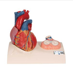 This model is cast from a real human heart and didactically prepared to facilitate a better understanding of the anatomy and blood flow of the heart. It shows the cardiac valves during diastole and on the base the valves are shown in systole. A dissection through the median plane makes an easy demonstration possible. Its attention to detail and high quality craftsmanship makes it definitely the top of the line heart model. The following feature set makes it stand out from the crowd and a must have for any health professional. All of the original heart structures were successfully obtained during the time consuming and detailed casting procedure making this model highly accurate and lifelike 2 atria and 2 ventricles show all the normal anatomical structures of the papillary muscles and heart valves Uniquely dissected in the median plane to optimally demonstrate the path of the oxygenated and deoxygenated blood The heart model shows both the diastolic and systolic state. In the model itself the valves are shown in the diastolic state and in the detail view on the base the valves are shown in the systolic state The heart valves are made of elasticated plastic making them very durable The base displays the heart in its natural position in the human body Life size cast from real human heart Easy and fun to use magnetic assembly (5 pieces) for easy demonstrations
3B Scientific Life-Size Human Heart Model, 5 Parts with Representation of Systole – 3B Smart Anatomy
-
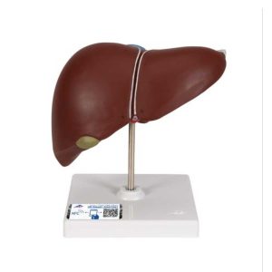 This realistic model shows the anatomy of the liver and gall bladder. The Liver with gall bladder shows: 4 lobes with gall bladder Extrahepatic ducts Hilus vessels This high quality liver with gallbladder replica is delivered on removable stand.
3B Scientific Liver Model with Gall Bladder
-
 This urinary system model shows the structures of the retroperitoneal cavity in the following details: Inferior vena cava Renal veins Aorta with its branches Iliacal vessels Ureter Urinary bladder Prostate Adrenal gland Rectum Musculature The right kidney of the male urinary system model is opened. This urinary system model has anatomical detail that makes it great for classroom or doctor’s office. Urinary system model is not delivered on base.
3B Scientific Male Urinary System Model, 3&4 Life-Size – 3B Smart Anatomy
-
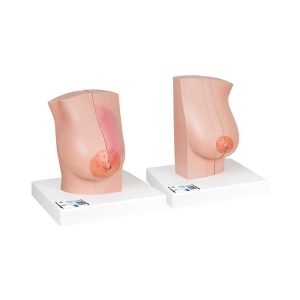 Use this model to teach your students about breast diseases, to educate your patients, and to raise awareness. The high quality of both the material used as well as the medically correct anatomy of the Female Breast Models make it a true 3B Scientific® product and a great educational tool for you. Both models are held together with magnets for easier demonstration. Lactating right breast: Medially divided into 2 halves Healthy lactating breath tissue on the cut surface of the external half Breast gland inflammation (mastitis) on the cut surface of the inner half Non-lactating left breast: 2 sagittal cuts, can be separated into 3 parts Healthy anatomical structures on the sectional plane of the external half Skin on the external half is windowed to view the regional lymph nodes Cysts and fibroadenoma on the external sectional plane of the middle breast cut Pathological proliferation of the breast connective tissue (fibrocystic breast disease) on the inner sectional plane of the middle breast cut Malignant tumors on the sectional plane of the inner breast cut
3B Scientific Model of Female Breast with Healthy and Unhealthy Tissue – 3B Smart Anatomy
-
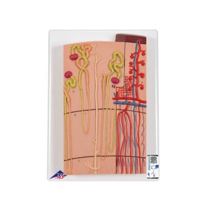 The nephron and blood vessel model is a nephron section depicting a section through a renal cortex and medulla. The nephron model features the renal corpuscles with proximal and distal convoluted tubules, loops of Henle, colleding tubules and blood vessels. The human nephron model is 120 times life size. The nephron and blood vessels model comes with a users manual as well. Nephrons and blood vessel model is mounted on a baseboard.
3B Scientific Nephrons and Blood Vessels Model, 120 times Full-Size – 3B Smart Anatomy
-
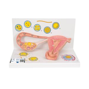 The model illustrates schematically how the ovum matures, how ovulation and fertilization occur and how the fertilized ovum develops to the stage where it embeds itself in the womb wall to begin the growth into an embryo. The various stages are shown in larger-than-life model form in an ovary, fallopian tube, and womb. An even more enlarged illustration of each is also printed on the base. Supplied on a base.
3B Scientific Ovaries and Fallopian Tubes Model with Stages of Fertilization, 2-times Magnified – 3B Smart Anatomy
-
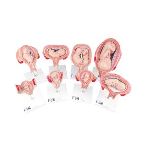 Our most popular series includes 8 models to show the complete stages of development. All models are mounted separately on a stand. 1st Month Embryo 2nd Month Embryo 3rd Month Embryo 4th Month Fetus (Transverse Lie) 5th Month Fetus (Breech Position) 5th Month Fetus (Transverse Lie) 5th Month Twin Fetuses (Normal Position) 7th Month Fetus Stands and uterus are separate and removable. In addition, the 4 largest fetuses can be removed from their uterus.
3B Scientific Pregnancy Models Series, 8 Individual Embryo and Fetus Models – 3B Smart Anatomy
-
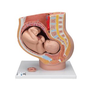 This anatomy model is a representation of a median section through the female pelvis at 40 weeks pregnant with a removable fetus. Study the normal position of child before birth with this model plus the human reproductive and urinary systems. A uterus with embryo in 3rd month of pregnancy is mounted on base for added detail. The realistic and high quality female pelvis includes the female genital organs and other important anatomical details. This pregnancy female pelvis is a great addition to any anatomy classroom or doctor’s office to educate about the stages of pregnancy.
3B Scientific Pregnancy Pelvis Model in Median Section with Removable Fetus (40 weeks), 3 part – 3B Smart Anatomy
-
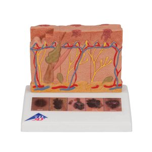 This 3B Scientific® Skin Pathology model shows healthy skin and 5 different stages of malignant melanoma on the front and back, enlarged 8 times: healthy malignant cells are found at the surface, within the epidermis malignant cells fill the epidermis, a few invade the papillary layer malignant cells fill the papillary layer malignant cells invade the reticular layer malignant cells have reached the subcutaneous fatty tissue, satellite cells approach a vein In the top view of the skin cancer model, the individual stages of externally visible skin changes are shown, allowing for an assessment according to the “ABCDE” criteria. The sides of the skin cancer model show the various levels of invasion into the skin layers according to Clark (I-V) and the tumor thickness according to Breslow (in mm). 5 original color illustrations on the base of the skin cancer model show various types of malignant melanomas. The skin cancer model comes mounted on a base. The skin cancer model is a great tool for illustrating this skin pathology.
3B Scientific Skin Cancer Model with 5 stages, 8 times magnified – 3B Smart Anatomy
-
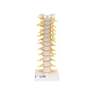 This spinal replica consists of the 12 thoracic vertebrae with intervertebral discs, thoracic nerves and spinal cord. This quality thoracic spinal column is affordable and anatomically correct. Spinal column delivered on flexible stand.
3B Scientific Thoracic Human Spinal Column Model
-
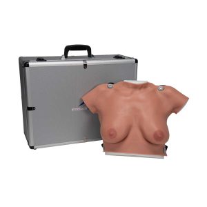 Demonstrate realistic self-examination with our natural casting of a female upper body with medium sized breasts. It can easily be worn, in order to better train and practice breast self-examination. • Made of new 3B SKINlike™ high-quality silicone • Displays the skin in finest detail • Very realistic to the touch and dermatologically tested • Breast examination is possible in both upright or lying positions • Benign and malignant tumors in different stages of development hone self-examination skills – 2 benign tumors – 4 malignant tumors – 2 typical anomalies • Includes “Female Breast” chart • Supplied with talcum powder, harness, stand and aluminium carrying case
3B Scientific Wearable Breast Self Examination Model – 3B Smart Anatomy
Zenrox Healthcare Solutions - Advanced Medical Equipment in Lagos
Zenrox offers cutting-edge medical equipment and comprehensive support services in Lagos. Explore our products designed for superior healthcare delivery. Contact us today for innovative medical solutions.