-
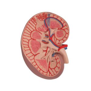 The kidney section, colorful and anatomically accurate, model depicts a longitudinal section of the human right kidney. All important structures of the human kidney for student and patient education are shown. The kidney is three times life size. No baseboard included with the basic kidney section.
3B Scientific Basic Kidney Section Model, 3 times Full-Size – 3B Smart Anatomy
-
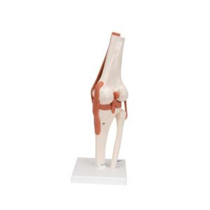 This high quality, life-size functional knee joint model clearly shows the anatomy and mechanics of the knee joint. This fully flexible knee joint model demonstrates abduction, anteversion, retroversion and internal/external rotation. The model consists of portion of femur, tibia and portion of fibula; also includes meniscus, patella with quadriceps tendon and joint ligaments, including the ACL and PCL. Delivered on removable stand for easy study or display.
3B Scientific Functional Human Knee Joint Model with Ligaments – 3B Smart Anatomy
-
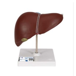 This realistic model shows the anatomy of the liver and gall bladder. The Liver with gall bladder shows: 4 lobes with gall bladder Extrahepatic ducts Hilus vessels This high quality liver with gallbladder replica is delivered on removable stand.
3B Scientific Liver Model with Gall Bladder
-
 This urinary system model shows the structures of the retroperitoneal cavity in the following details: Inferior vena cava Renal veins Aorta with its branches Iliacal vessels Ureter Urinary bladder Prostate Adrenal gland Rectum Musculature The right kidney of the male urinary system model is opened. This urinary system model has anatomical detail that makes it great for classroom or doctor’s office. Urinary system model is not delivered on base.
3B Scientific Male Urinary System Model, 3&4 Life-Size – 3B Smart Anatomy
-
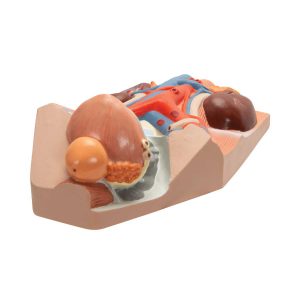
 The nephron and blood vessel model is a nephron section depicting a section through a renal cortex and medulla. The nephron model features the renal corpuscles with proximal and distal convoluted tubules, loops of Henle, colleding tubules and blood vessels. The human nephron model is 120 times life size. The nephron and blood vessels model comes with a users manual as well. Nephrons and blood vessel model is mounted on a baseboard.
3B Scientific Nephrons and Blood Vessels Model, 120 times Full-Size – 3B Smart Anatomy
-
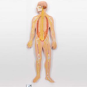 This nervous system relief model shows a schematic representation of the central and peripheral nervous system. An excellent nervous system model to study the structure of the human nervous system. This nervous system model is 1/2 life size and is a great teaching tool. The nervous system model is delivered on baseboard.
3B Scientific Human Nervous System Model, 1-2 Life-Size – 3B Smart Anatomy
-
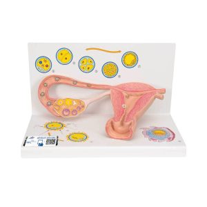 The model illustrates schematically how the ovum matures, how ovulation and fertilization occur and how the fertilized ovum develops to the stage where it embeds itself in the womb wall to begin the growth into an embryo. The various stages are shown in larger-than-life model form in an ovary, fallopian tube, and womb. An even more enlarged illustration of each is also printed on the base. Supplied on a base.
3B Scientific Ovaries and Fallopian Tubes Model with Stages of Fertilization, 2-times Magnified – 3B Smart Anatomy
-
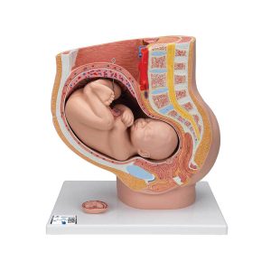 This anatomy model is a representation of a median section through the female pelvis at 40 weeks pregnant with a removable fetus. Study the normal position of child before birth with this model plus the human reproductive and urinary systems. A uterus with embryo in 3rd month of pregnancy is mounted on base for added detail. The realistic and high quality female pelvis includes the female genital organs and other important anatomical details. This pregnancy female pelvis is a great addition to any anatomy classroom or doctor’s office to educate about the stages of pregnancy.
3B Scientific Pregnancy Pelvis Model in Median Section with Removable Fetus (40 weeks), 3 part – 3B Smart Anatomy
-
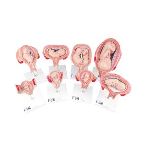 Our most popular series includes 8 models to show the complete stages of development. All models are mounted separately on a stand. 1st Month Embryo 2nd Month Embryo 3rd Month Embryo 4th Month Fetus (Transverse Lie) 5th Month Fetus (Breech Position) 5th Month Fetus (Transverse Lie) 5th Month Twin Fetuses (Normal Position) 7th Month Fetus Stands and uterus are separate and removable. In addition, the 4 largest fetuses can be removed from their uterus.
3B Scientific Pregnancy Models Series, 8 Individual Embryo and Fetus Models – 3B Smart Anatomy
-
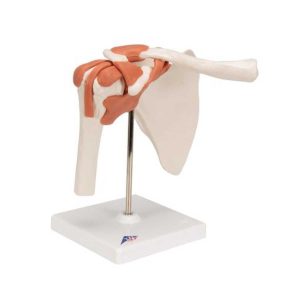 This high quality life-size functional shoulder joint model shows the anatomy and mechanics of the shoulder joint. Consisting of the scapula, clavical, portion of humerus and joint ligaments, this fully flexible shoulder joint model clearly demonstrates abduction, anteversion, retroversion and internal/external rotation. The Functional shoulder joint model comes on a stand for easy study and display.
3B Scientific Functional Human Shoulder Joint – 3B Smart Anatomy
-
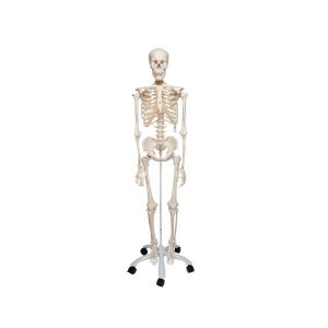
 Fred offers all the advantages of a 3B Scientific® skeleton. Fred’s bendy spine can copy almost any human movement too. Once bent, Fred will remain in that position, to show correct and incorrect postures as well as any pathological malalignments. All skull movements can be shown on the head joints. Protruding spinal nerves and vertebral arteries are also shown on this skeleton, as well as a dorsolateral slipped disc between the 3rd and 4th lumbar vertebrae. Now available on a stable metal stand with 5 casters! Here are Fred’s other advantages: • Top quality, life-size natural casting • Made from a durable, unbreakable synthetic material • 3-part assembled skull with individually inserted teeth • Close to the realistic weight of around 200 bones • Easy to remove limbs • On a stable metal stand with 5 casters (painted white) • Exceptional value for money • Final assembly carried out by hand • 3 year guarantee Comes with metal stand and transparent dust cover.
3B Scientific Flexible Human Skeleton Model Fred – 3B Smart Anatomy
-
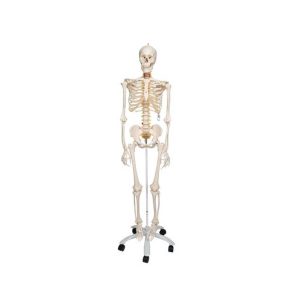
 Proven Quality – even more stable! Stan, the standard model of a human skeleton, has been appreciated throughout the world for decades. Thanks to its very high quality and robust construction, it is perfect for use in hospitals, schools, universities and laboratories. The other advantages of the 3B Scientific® skeleton are: • Exceptional value for money • 3 year guarantee • Top quality natural casting • Final assembly carried out by hand • Made from a durable, unbreakable synthetic material • On a stable metal stand with 5 casters (painted white) • Close to the realistic weight of around 200 bones • Natural skeleton size • 3 part assembled skull • Individually inserted teeth • Limbs can be removed quickly and easily • Skull with magnetic connections Comes with metal stand and transparent dust cover.
3B Scientific Human Skeleton Model Stan – 3B Smart Anatomy
-
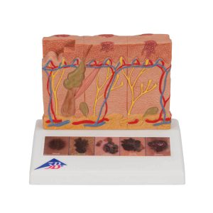 This 3B Scientific® Skin Pathology model shows healthy skin and 5 different stages of malignant melanoma on the front and back, enlarged 8 times: healthy malignant cells are found at the surface, within the epidermis malignant cells fill the epidermis, a few invade the papillary layer malignant cells fill the papillary layer malignant cells invade the reticular layer malignant cells have reached the subcutaneous fatty tissue, satellite cells approach a vein In the top view of the skin cancer model, the individual stages of externally visible skin changes are shown, allowing for an assessment according to the “ABCDE” criteria. The sides of the skin cancer model show the various levels of invasion into the skin layers according to Clark (I-V) and the tumor thickness according to Breslow (in mm). 5 original color illustrations on the base of the skin cancer model show various types of malignant melanomas. The skin cancer model comes mounted on a base. The skin cancer model is a great tool for illustrating this skin pathology.
3B Scientific Skin Cancer Model with 5 stages, 8 times magnified – 3B Smart Anatomy
-
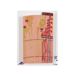
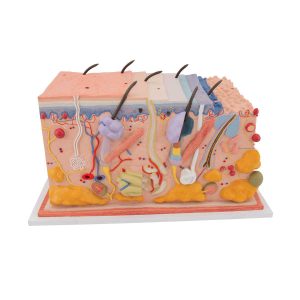 This unique skin model shows a section of human skin in three dimensional form. Individual skin layers are differentiated, and important structures of the skin such as hair, sebaceous and sweat glands, receptors, nerves and vessels are shown in detail. The high quality skin block model is mounted on baseboard. Demonstrating the anatomy of the human skin has never been easier! This skin block model details the human skin in 70 times life size.
3B Scientific Human Skin Section Model, 70 times Full-Size – 3B Smart Anatomy
-
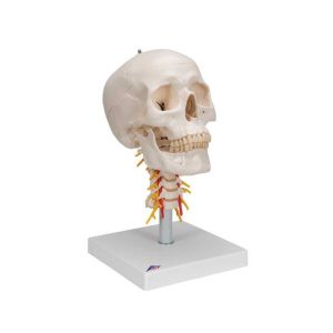 This human skull replica is flexibly mounted on a cervical spine. Features also shown are the hindbrain, spinal cord, cervical nerves, vertebral arteries, basilar artery and rear cerebral arteries. Skull can be removed from stand. High quality original human skull cast Skull is handmade from hard, unbreakable plastic Highly accurate representation of the fissures, foramina, processes, sutures etc. Can be disassembled into skull cap, base of skull and mandible Mandible of skull is mounted on a spring to easily demonstrate natural movement Add this detailed human skull model to your collection today!
3B Scientific Human Skull Model on Cervical Spine, 4 part – 3B Smart Anatomy
-
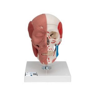 This model of the face musculature by 3B Scientific is used to easily demonstrate the causes of temporomandibular disorders and other dysfunctional disturbances of the TMJ and masticatory muscles. The face and mastication muscles are illustrated on the right half of this skull and the face musculature can easily and precisely be differentiated from the mastication musculature by using two colors. On the left half the muscle origins and insertions are marked with colors as well(origin: red, insertion: blue). The jaw is movable and due to the flexible musculature the rudimentary chewing motion can be demonstrated. Cranium and m. masseter are easily detachable. Only the highest quality material was used to manufacture this made in Germany model. Its durability makes it perfect for hands-on teaching. Whether it is used in a classroom or a doctor‘s office, the demonstration is as realistic as a model can be. Now with magnetic connections.
3B Scientific Human Skull with Facial Muscles – 3B Smart Anatomy
Zenrox Healthcare Solutions - Advanced Medical Equipment in Lagos
Zenrox offers cutting-edge medical equipment and comprehensive support services in Lagos. Explore our products designed for superior healthcare delivery. Contact us today for innovative medical solutions.