-
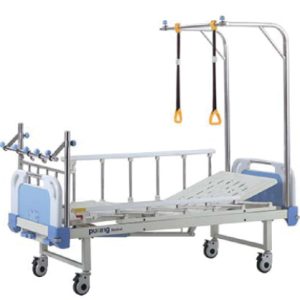 Epoxy coated bed frame, with detachable lat tube headboards 2 function, which can be operated by crank, the details are as follows: back rest: 0º-70º±2º : knee rest: 0º-35º±2º with stainless steel traction frame at the foot/head end. Anti-bumper in four corners, with I.V. rod locations Anti-noise rubber at the foot base Freight saving knock-down construction.
Full-fowler orthopedics bed C-5-1
-
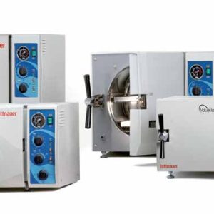 EA / EKA – Fully AutomaticAutoclaves Closed Door Active Drying Air Pump: With extra fast and efficient drying cycles, the EKA and EA autoclaves significantly increase your productivity. These two models have Model Chamber Volume Cold Cycle Time Hot Cycle Time EA Series 2340EA 2540EA 3850EA 3870EA 19Liter 23Liter 64Liter 85Liter 23min. 25min. 31min. 31min. 16min. 18min. 20min. 20min. EKA Series 2340EKA 2540EKA 19Liter 23Liter 14min. 14min. 11 min. 11 min. the added benefit of a high efficiency air pump which allows closed door active drying. The EKA and EA are built for improved sterilization with the ability to dry packs and pouches. Benefits: More thorough drying and sterilization Faster drying for a shorter overall cycle 0.2 µm HEPA air filter provides sterile, bacteria-free air for drying Tested for unwrapped instruments. Cycle times includes heat up, sterilization exposure and exhaust. All cycle times may vary with instrument load and voltage.
Fully Automatic Autoclaves
-
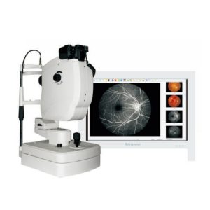 Fundus Camera(SK-650A) is a non mydriatic fundus photography machine mainly used for retina diseases examination. You can get high resolution pictures with this fundus camera. Professional software will be offered, and you can easily manage patient information, process pictures, and generate analysis reports. It has Dicom 3.0 interface that can be connected to HIS/PACS system. Features: Test Mode: Non Mydriatic Fundus Photography Red Free Photography: Yes Digital Camera: Canon EOS Technology Color Image Resolution: 5184px*3456px Minimum Pupil Size: 3.3mm Diopter Compensation: -25D~+30D Max Angle of View: 53° Working Distance: 40mm±2mm
Fundus Camera
-
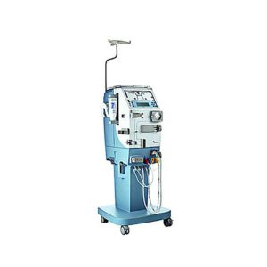 Features: The ultimate value of perfecting performance. As the leading innovator in renal care, Gambro is constantly seeking new ways to perfect the performance of therapy delivery. The compact AK 96 machine enables dialysis providers to consistently deliver the highest level of quality and safety in hemodialysis (HD) treatment with improved cost-efficiency. Innovative features such as the Diascan system and a completely new user interface offer a simpler way to consistently deliver flexible high quality, treatments for patients. Specifications: Model Name/Number: AK96 Application: Haemodialysis Power: 240v Accuracy: High Display Range: LED Display Body Material: PVC Brand: Gambro Types Of Dialysis Machine: Clinical Use Operation Mode: Automatic Model: AK96 Click here To Download Catalogue
Gambro Dialysis Machine
-
Hot
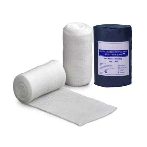
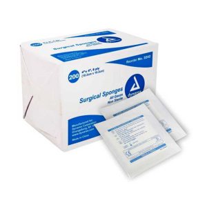 Part of every first-aid kit should be some gauze sponges in various sizes. The sponges are useful in cleaning wounds and dressing them as well as many other things. The Dynarex® Non-Sterile Gauze Sponges – 2″ x 2″ make a great addition to any doctor’s office, nurse’s station or home first-aid kit. Nonstick gauze 2″ x 2″ 5000 sponges per case Cotton field squares Absorbent dressing
Gauze Pad
-
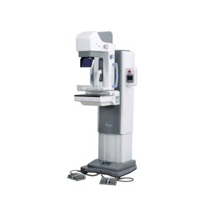
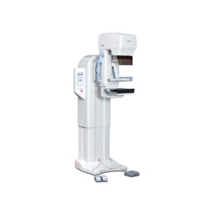 MX-600 is designed compactly for easy install and operation. Features: Auto Standard Positioning system (ASP) With Auto Standard exposure Positioning systems designed to maximize the convenience of radiography, you can easily adjust positioning by using standard exposure (RCC, LCC, RMLO, LMLO). ASP function makes operators easy to execute 4 axis exposures by software programming. One-touch button controls 4 standard positions (RCC, LCC, RMLO, LMLO) using C-Arm pre-set adjustment (MedioLaterral, Oblique view and CranioCaudal view). The ISO level can adjust the level of MX-600 standing position when it operates from vertical exposure to oblique and vice versa. Intelligent Automatic Exposure Control (AEC) Automatic Exposure Control system enables production of images with reliable intensity for any film, screen, or digital radiography. Furthermore, it greatly enhances the convenience of radiography by embedding the Full-AEC function which utilizes Auto kV control. It sets the best diagnosis environment by reducing manual operation and increasing patient throughput. Compression with comfort in mind When pressure is required for Mammography, it allows you to apply appropriate amount of pressure (up to maximum of 20kg) and is equipped with MICOM control’s Soft-touch system which minimize the discomfort. Automatic Conversion (Filter) Motorized and manual breast compressions are available for MX-600. MX-600 displays compression level and thickness on main body’s panel. Stable Output A molybdenum (0.03mm Mo) and Aluminum (0.5mm Al) filter are installed to absorb unnecessary x-ray. Mo filter covers low level kV range (22-35kV) and Al filter covers high kV range (35-39kV). Mo filter is useful for increasing image contrast in large breast with large amounts of glandular tissue. The rhodium filter (0.025mm Rh) can be installed as an option (as replacement of Al filter). Display of Exposure Condition When some degree of pressure is required for radiography, it allows you to apply the appropriate pressure (up to a maximum of 20kg) and is equipped with MICOM control’s Soft-touch system which is designed to minimize the discomfort of the examine with in the pressure range. Technical Specifications: Generator Type: High frequency inverter (40 kHz). Radiographic qualification: Great focus 22-39kV / 1-600mAs. Small bulb 22-35kV / 1-100mAs. Tube Focal point size: 0.1 / 0.3mm. Anode heat capacity: 300KHU (molybdenum). Filtration: Mo. Radiographic support Movement up / down: 720-1420mm (motorized). Rotating movement: ± 180˚ (Motorized) . Automatic standard positioning (ASP). SID: 650 mm. Bucky device Cassette size: 18x24cm, Grid: 4: 1, 91 lines / inches. Automatic exposure control (AEC) Type: solid state detector. Mode: 3 modes (Full / Semi / Manual). Density adjustment: 19 steps. Collimator: Automatic. Accessories Face protection. Film marker, hand switch, point compression paddle. 18x24cm Bucky device with compression paddle. Collimating device (18×24 and dotted plate). Options Set of Bucky devices of 24×30 cm. Set of 1.5X or 1.8X magnification devices. Click Here To Download Catalogue
Genoray Analogue Mammography MX-600
-
 The DMX-600 is a full-field digital mammography system with smart C-arm rotation and fully motorized movements for fast and accurate positioning and efficient examinations. It features crystalline Silicon CMOS active pixel detector for higher contrast and higher resolution images and dual target X-ray tube for dose reduction. Features: Excellent & High resolution Image quality: – Innovative and accurate technology of Crystalline Silicon CMOS active pixel detector with higher contrast, higher resolution, brilliant images and economic maintenance cost. – High performance of dual target X-ray tube for dose reduction. – High output power of HF Generator with stability and efficiency. – Stable hardware with the best reproducibility. Large F.O.V. (Field Of View) by Multi-format technology: – Optimally suited for screening and diagnosis with the financial advantages. – Patent Application No. : 10-2011-0139676 – Available large F.O.V. to 23x23cm for almost patient. User friendly Versatile functions: – Smart C-arm rotation and fully motorized movements for fast & accurate positioning and efficiency examination by ASP & ASE (CC, RMLO, LMLO) – Smart Automatic collimation with easy operation. – Smart compression System by Microprocessor and Intelligent AEC control. – Ergonomic design for quick and easy set-up. Smart Software user alike: – Customized and dedicated acquisition workstation. – Perfect PACS accessibility with full DICOM capability. – Extremely fast saving and transferring images for quick access and optimized workflow. Click Here To Download Catalogue
Genoray Full Field Digital Mammography DMX-600
-
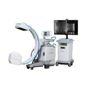 The OSCAR 15 is a culmination of several years worth of developmental experience from Genoray. With CMOS imaging excellence & 15kW HFG you diagnosis need will be met while improving your productivity especially DSA (Digital Subtraction Angiography). APPLICATION General Surgery Office based Vascular Center Pain Management Orthopdics Urology Cardiac Procedures Hybrid OR Neuro & Spine Surgery Pain Management Orthopedic Surgery Trauma Procedure -Urology Procedure Cardiac Surgery Peripheral Artery Diseases -Vascular Surgery SPECIFICATION 260 x 260 mm CMOS Type Flat-Panel detector for distortion-free imaging (High resolution images, Wide FOV, Low Noise) 15kW HFG 4″Touch LCD monitor 43″ LCD Monitor Dual Foot Switch DICOM 3.0 -CD/DVD Burner – USB Port 800mm free space and 150° (+90°,-60°) orbital rotation, SID 1,000mm 155º Dynamic Orbital Rotation 2 kW Stationary Anode X-Ray Tube 5 Million Images Storage Capacity FEATURE Exceptional Image Quality With an optimal flat panel detector size of 26 x 26cm you won’t miss a thing with its high quality resolution. It makes accurate diagnosis in a variety of departments especially DSA Low Dose Mode Low dose mode is desgined to acquire reasonable image to diagnose the patient with minimum dosage. Edge Enhancenment For user to get more accurate diagnosis result enhancing edge of image. Motion Correction This function detects the movement and reduce the after image while exposing X-ray. Metal Correction To prevent over-dose radiation or low quality image casued by metal inturrption on field of view. Virtual Collimator The virtual collimator allows for the selection of your desired field of view, while reducing the amount of radiation exposure by limiting the X-Ray beam. Auto Collimation Prevention of unncessary X-Ray exposure by focusing on the area of interest while autmoatically collimating the remaining areas. POWERFULL SOFTWARE ZENIS A total solution from acquisition, storage, management, communication to print out. Provide convenient environment from user-centric interface. Diagnosis and confirm from recognizable simple icons. Convenience of database management. Convenient diagnostic functions for easy patient / image management Accurate diagnostic tools Improve the efficiency of your hospital management Perfect compatibility with all PACS A must have for a digitally equipped hospital Convenient communication and management for your customers Dicom Support DIGITAL SUBTRACTION ANGIOGRAPHY Native DSA Pairing fluoroscopy with constrast media to display the basic angiography views Motion Matching Selects the proper mask to apply and remove artifacts made by a patient’s movement or breathing Post-Processing Processing: Improvement of the processed image after the DSA procedure Landmarking / Brightness / Contrast After setting the position for a vessel, the subject can be placed back to their original position by using the shift function to compensate for any movement. Allows for various functions that assist with accurately inserting a catheter. Peak Opacification Ability to diagnose a blood vessel with only a small amount of contrast media Road Mapping, Land Mark After setting a position for a vessel, the subject can be moved back to their original place by using the shift function to compensate for any movement. Provides various functions that helps accurately to insert a guide wire, catheter is compatible with the hybrid operating room. Auto Roadmap Mask Obtain blood vessel type information while only using a small amount of contrast media Manual Roadmap Mask Roadmap your vessels using a prevoiusly taken DSA image Roadmap Pixel Shift Re-position the roadmap mask by shifting the pixels to the proper position Click Here To Download Catalogue
Genoray OSCAR 15 Surgical C-Arm Machine
Zenrox Healthcare Solutions - Advanced Medical Equipment in Lagos
Zenrox offers cutting-edge medical equipment and comprehensive support services in Lagos. Explore our products designed for superior healthcare delivery. Contact us today for innovative medical solutions.