-
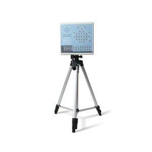 KT88-3200 Digital Brain Electric Activity Mapping collects EEG signal with electrodes, via integrated amplification, A/D transformation, PC auto-analysis, FFT, to form electroencephalogram that displays with color depth. The product is applicable for checking such diseases as epilepsy, intracranial inflammation, cerebrovascular diseases and brain tumors. Features: Method of 10/20 electrodes placement under international standard system, leads can be changed during replaying. Support different types of combined leads during sampling. Adopt bioelectricity amplifier for distilling brainwave, continuous recording time can be up to 24 hours, integrated full-automatic calibration system. Powerful playback function: amplitude and display speed are adjustable. The special subdividing time line divides the waveform in one second into 5 parts, which is easy for doctors to look over the waveform. Digital filter system can be set as required, providing different window types EEG signal clipping function, analyze and store any section of EEG wave, and select several waveform segments for automatically analyzing and distilling to different parameters. Electronic frequency ruler, convenient to measure the basic information of any appointed EEG waveform. With partial enlarging window, accurate measurement of EEG period, amplitude and frequency, which can be adjusted according to personnel judgment. Mark EEG wave under the events of opening eyes, closing eyes and flashing with different colors, and user-defined events can be added, waveform color for evoked event can also be set freely, ensures that the waveform in corresponding time can be rapidly found by event name during case playback. Powerful automatic analysis function, can carry through the power spectrum analysis and pathologic wave detection for appointed waveform. Many graphs can be displayed in the same screen, including kinds of BEAM, numerical BEAM, compressed spectrum graph, trend graph, and so on. Professional isolation transformer, dual power supply isolation system and optoelectronic data transmission to ensure security. Use USB interface to transmit data which just need to be inserted. Multifunctional flash stimulator of USB interface, and flashing can be controlled manually or automatically. A flash stimulation scheme can be set and performed in the process of sampling. Perfect case management function, provides many means for research and quick statistic information; convenient case export and import function, and stores with MO or CD-RW disk, which is easy for data research. Integrative image and character report, report can be edited in mode and switched to Word document. Case files can be transformed into EDF and BDF data format, convenient for data exchange, academic exchange and further analysis. System parameters and display modes can be set as required, which meets different user’s requirement. Add marks and annotations to the waveform designated, which can rapidly find the waveform in that time by marks. Optional video function: USB camera is easy to install, convenient to use and exact to record. With flexible playback function, which can browse the waveform of any time along with the corresponding isochronous sampled image. SpO2 function is optional. Technical Specifications: 32 channels of EEG Sampling rate: 200 dots/s Accuracy: 12bit Input impedance: ≥10MΩ Patient leak current: < 10µA Noise level: ≤5µVp-p CMRR: ≥90dB Magnification multiple: 10000 Filter constant: all digital and free enactment Display speed(paper speed): 5, 10, 15, 30, 60, 120 mm/s Amplitude: 1, 1.5, 2, 3, 5, 7.5, 10, 12, 15, 20, 30, 50 mm/50µV Playback speed:1 time, 2 times, 3 times, 10 times, 20 times, 40 times, 60 times 50Hz interference suppression: ≥30dB Safety type:Class II, type BF applied part Accesories: EEG lead EEG electrode Data line Headgear Strobe light Strobe light power supply PC software CD User Manual Earth wire EEG bracket Aluminum block Click Here To Download Catalogue
Contec KT88-3200 Electroencephalogram (EEG) Machine
-
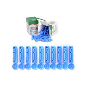
 Deep vein thrombosis DVT-7700 MAIN is a DVT machine with HOSE and Accessories (Disposable sleeve thigh(M), Disposable sleeve calf(M), Disposable sleeve foot, and Tube (pair)) – Deep vein thrombosis (DVT) happens when a blood clot forms in a vein deep inside the body. Veins in the thigh, foot, and calf create a natural pumping system that helps flow blood back to the heart. But when an individual is immobile, blood circulation is decreased, and the risk of developing a blood clot goes up. DVT machine Specifications: Power requiremnet: 100V~240VAC, 50/60Hz Power Consumption: Maximum 18w Power Cord: Hospital Grade Plug (Detachable) Operation Mode: A1,A2,A3,B1,B2,B3,B4,C1,D1,D2 Adjustable Timer: 0-99 Hours Applied Pressure Leg sleeve: 30~60 mmHg (Caif, Thigh) Food Cuffs: 130mmHg Operation Mood: Continuous Compression Type: Leg sleeve: Gradient, Sequential Food Cuffs: Uniform Dimension: 200mm (L) * 190mm (W) * 260 mm (H) Weight: 2.3 kg (5.07 ib) Battery: Capacity 10.8 Vdc 4,400 mAh Lithiumion (optional) Run Time 6-8 Hours Click Here To Download Catalogue
Deep Vein Thrombosis DVT-7700
-
Hot
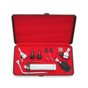 Features: Consisting of Otoscope 3 Speculae Nasal Speculum Bent Arm Illuminator Tongue Depressor Laryngeal Mirror Battery
Diagnostic Set
-
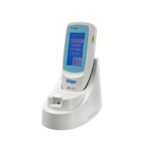 Dräger Jaundice Meter JM-105 is a non-invasive transcutaneous bilirubinometer that measures the yellowness of subcutaneous tissue in newborn infants as young as 24 weeks gestational age who have not undergone a transfusion or phototherapy. The Dräger Jaundice Meter JM-105 gives you consistent quality, cost effectively delivered over the lifetime of the device. Features: Effective Jaundice Screening With the Dräger Jaundice Meter JM-105 you can accurately identify at-risk infants as young as 35 weeks gestational age. Effective screening can decrease readmission rates and durations of stay. Having dependable results in seconds rather than hours increases patient safety and expedites decision making. New integrated flagging feature helps you keep track of patients in need of special attention and comply with your jaundice management protocols. Easy to use, easy on everyone The Dräger Jaundice Meter JM-105 expands the concept of ease of use. It also means an easier experience for all involved. Since there’s no blood draw, there’s less stress on fragile new-borns and their parents – which makes things easier on you. Screening with this device is fast and easy; simply clean the reusable tip with an alcohol wipe and take your measurement. No fumbling with disposable tips. No recalibration with every baby. Finally, the device makes it easy for you to get the information you need because it eliminates waiting for laboratory results. And because the device connects to your hospital information system, transferring jaundice screening information to the infant’s electronic medical record is effortless and accurate. Simply put, the device is gentle for the new-born and efficient for you. Improved process and cost efficiency The Dräger Jaundice Meter JM-105 streamlines jaundice screening practices by eliminating time-consuming blood draws, scheduling of laboratory work, and processing costs. It improves efficiency by delivering reliable measurements with significantly fewer steps, which frees up more time for direct infant care. The data transfer functionality and barcode scanner help you optimise your screening program and reduce the risk of human error. Because it has a reusable probe tip, it requires no disposables. With the volume of screenings performed in nurseries today, the cost of such disposable items can ultimately exceed the initial cost of the device itself. As a result, you save time and money while delivering an exceptional standard of care with Dräger Jaundice Meter JM-105 Click Here To Download Catalogue
Dräger Jaundice Meter JM-105
-
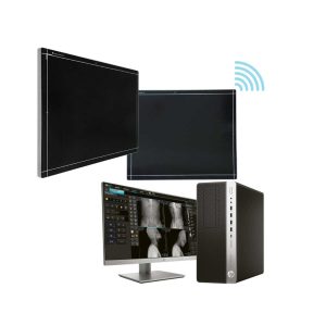 DRGEM ACQUIDR (DRGEM DR System) is the digital imaging system composed of a Flat Panel Detector(FPD) and an imaging workstation with software. The digital FPD and full-feature imaging software with excellent digital image processing will meet all your needs in the diagnostic digital radiographic field. Features of DRGEM DR System: Adaptable with most existing analog x-ray systems AED(Auto Exposure Detection) function available Simple and convenient installation Intuitive graphic user interface Over 1300 pre-loaded exam profiles with DRGEM’s generator Configurable automatic study PMS / EMR /HIS / RIS work list support Multiple printing formats DICOM 3.0 compliant Brightness and contrast adjustment Rotation Horizontal and vertical flip Zoom and pan Left / right makers Optimized work flow Automatic shutters Support DAP interface Exposure index (EI) DR upgrade solution by retrofit. Portable and Wireless FPD DR solution for maximum flexibility in virtually all general radiographic specialties. Portable and wireless detector fits into most existing analogue x-ray system. Faster workflow after DR upgrade. With AED(Auto Exposure Detection) function, there is no DR trigger cable between detector and generator providing very easy DR upgrade. Easy to interface with any kind of x-ray generator. Radmax acquisition workstation Turns any analogue X-Ray system into a fully DR system Optional image stitching program Vet software available Detector format: 17×17” / 17×14″, wired/wireless
DRGEM DR System
-
Hot
 Features: ●Professional Same ear wax removal tool as those used by doctors, you can easily eliminate ear wax buildup at home, really save your money and time on medical visiting. Safe and Environmentally Friendly. ●Quick & Easy This ear wax removal kit is a quick, effective treatment for excess ear wax buildup. Fill the bottle with solution, Twist on the disposable tip, Use the trigger handle to spray solution into the ear canal. So Easy. ●Standard Capacity of the ear cleaner solution bottle is 10.6Oz, it has the most suitable size to hold in hand. Working at condition 32-122℉(0-50℃). Recommend to fill 1/5 of the bottle with OTC hydrogen peroxide, and 4/5 with very warm water. ●Complete Ear Washer System Our earwax removal kit comes with 1× Ear Washer Bottle, 1× Wash Basin, 1× Rubber Bulb, 1× Short Injection Head, 1× Long Hose Injection Head, 5× Disposable Tip, 1× User Manual.
Ear Irrigation and acumen removal
-
Hot
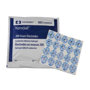 The Covidien/Kendall 200 series electrodes are a cost effective general adult monitoring electrode. The foam substrate repels fluids and conforms to body contours. The Covidien/Kendall™ 200 series electrodes have a smaller footprint for convenient lead placement and a teardrop shape for easy removal from the liner and patient. Many packaging configurations are offered to satisfy all customer needs. The patented conductive adhesive hydrogel is designed to stay fresh up to 45 days out of the package. Features: Smaller, Foam ECG monitoring electrodes with Covidien’s patented adhesive hydrogel Proprietary Conductive Adhesive Hydrogel provides additional adhesion, consistent tracings, no messy residue, non-irritating and stays fresh up to 45 days out of the package High Quality Foam resists fluids and conforms easily to skin to ensure excellent trace quality Smaller Size allows for easy lead placement on smaller patients Tear Drop Shape & Lift Tab facilitates easy removal from release liner and patient Non-irritating Gel Formula patient comfort and reduced clinician time Size: 1.5″ diameter 10 ECG Electrodes per pack, 50 packs per case Sold by the case (500 electrodes) Manufacturer: Covidien Manufacturer’s Product ID: 31499224
ECG Electrodes
-
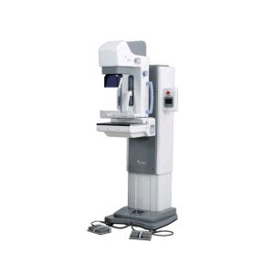
 MX-600 is designed compactly for easy install and operation. Features: Auto Standard Positioning system (ASP) With Auto Standard exposure Positioning systems designed to maximize the convenience of radiography, you can easily adjust positioning by using standard exposure (RCC, LCC, RMLO, LMLO). ASP function makes operators easy to execute 4 axis exposures by software programming. One-touch button controls 4 standard positions (RCC, LCC, RMLO, LMLO) using C-Arm pre-set adjustment (MedioLaterral, Oblique view and CranioCaudal view). The ISO level can adjust the level of MX-600 standing position when it operates from vertical exposure to oblique and vice versa. Intelligent Automatic Exposure Control (AEC) Automatic Exposure Control system enables production of images with reliable intensity for any film, screen, or digital radiography. Furthermore, it greatly enhances the convenience of radiography by embedding the Full-AEC function which utilizes Auto kV control. It sets the best diagnosis environment by reducing manual operation and increasing patient throughput. Compression with comfort in mind When pressure is required for Mammography, it allows you to apply appropriate amount of pressure (up to maximum of 20kg) and is equipped with MICOM control’s Soft-touch system which minimize the discomfort. Automatic Conversion (Filter) Motorized and manual breast compressions are available for MX-600. MX-600 displays compression level and thickness on main body’s panel. Stable Output A molybdenum (0.03mm Mo) and Aluminum (0.5mm Al) filter are installed to absorb unnecessary x-ray. Mo filter covers low level kV range (22-35kV) and Al filter covers high kV range (35-39kV). Mo filter is useful for increasing image contrast in large breast with large amounts of glandular tissue. The rhodium filter (0.025mm Rh) can be installed as an option (as replacement of Al filter). Display of Exposure Condition When some degree of pressure is required for radiography, it allows you to apply the appropriate pressure (up to a maximum of 20kg) and is equipped with MICOM control’s Soft-touch system which is designed to minimize the discomfort of the examine with in the pressure range. Technical Specifications: Generator Type: High frequency inverter (40 kHz). Radiographic qualification: Great focus 22-39kV / 1-600mAs. Small bulb 22-35kV / 1-100mAs. Tube Focal point size: 0.1 / 0.3mm. Anode heat capacity: 300KHU (molybdenum). Filtration: Mo. Radiographic support Movement up / down: 720-1420mm (motorized). Rotating movement: ± 180˚ (Motorized) . Automatic standard positioning (ASP). SID: 650 mm. Bucky device Cassette size: 18x24cm, Grid: 4: 1, 91 lines / inches. Automatic exposure control (AEC) Type: solid state detector. Mode: 3 modes (Full / Semi / Manual). Density adjustment: 19 steps. Collimator: Automatic. Accessories Face protection. Film marker, hand switch, point compression paddle. 18x24cm Bucky device with compression paddle. Collimating device (18×24 and dotted plate). Options Set of Bucky devices of 24×30 cm. Set of 1.5X or 1.8X magnification devices. Click Here To Download Catalogue
Genoray Analogue Mammography MX-600
-
 The DMX-600 is a full-field digital mammography system with smart C-arm rotation and fully motorized movements for fast and accurate positioning and efficient examinations. It features crystalline Silicon CMOS active pixel detector for higher contrast and higher resolution images and dual target X-ray tube for dose reduction. Features: Excellent & High resolution Image quality: – Innovative and accurate technology of Crystalline Silicon CMOS active pixel detector with higher contrast, higher resolution, brilliant images and economic maintenance cost. – High performance of dual target X-ray tube for dose reduction. – High output power of HF Generator with stability and efficiency. – Stable hardware with the best reproducibility. Large F.O.V. (Field Of View) by Multi-format technology: – Optimally suited for screening and diagnosis with the financial advantages. – Patent Application No. : 10-2011-0139676 – Available large F.O.V. to 23x23cm for almost patient. User friendly Versatile functions: – Smart C-arm rotation and fully motorized movements for fast & accurate positioning and efficiency examination by ASP & ASE (CC, RMLO, LMLO) – Smart Automatic collimation with easy operation. – Smart compression System by Microprocessor and Intelligent AEC control. – Ergonomic design for quick and easy set-up. Smart Software user alike: – Customized and dedicated acquisition workstation. – Perfect PACS accessibility with full DICOM capability. – Extremely fast saving and transferring images for quick access and optimized workflow. Click Here To Download Catalogue
Genoray Full Field Digital Mammography DMX-600
-
Hot
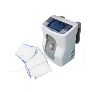
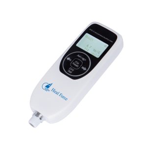 Jaundice is one of the most common symptoms in the neonatal period. Clinical reports show that 20% to 30% of newborns may have visible jaundice. And early neonatal pathological jaundice is prone to bilirubin encephalopathy, so it is important to screen for early pathological jaundice. DHD-C Percutaneous Jaundice Meter physical sample and advantages Large LCD display Two units displayed at the same time Average value display, more storage Lithium battery, long standby time Continuous measurement, no need to reset Technical Parameters: Measurement meth: light reflective Light source: LED Display: LCD Indication error: 0~25 mg/ dL ± 1.0 mg/ dL Power supply: Lithium battery (DC3.6V, 1000mAh ± 20%);Tests can be done more than 800 times after a fully charge; Calibration color screen: white end face is 0, yellow end face is 16.0±1.0 Function Simultaneous display of measurement units: displaying mg / dL & µmol / L in the meantime. Data Storage: 200 groups of measurement data Average Calculation: It can measure the average value of 2-5 times Start-up preparation time: Ready to use at startup, no preparation required Battery voltage detection function: When the battery voltage of the tester is too low, the screen displays “Low Battery” Automatic shutdown: Automatic shutdown after 10 minutes of non-working state Battery display: Real-time display of battery power Charging display: The viewing screen shows “Charging … ” during charging. Automatic charging protection: It will stop charging automatically at the time battery voltage charging arrive 4.2V±0.05V. Click Here To Download Catalogue
Heal Force Percutaneous Jaundice Meter DHD-C
-
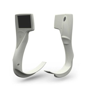 Features: i-view is the new, single use, fully disposable video laryngoscope providing the option of video laryngoscopy wherever you might need to intubate. It offers a cost effective solution, by combining all the advantages of a fully integrated video laryngoscope in a single use, disposable product. i-view incorporates a Macintosh blade, so can also be used for direct as well as video laryngoscopy. The ergonomic design ensures i-view is easy and instinctive to use and the integral LCD screen provides an optimal view in a variety of light conditions; and it’s ready for use seconds after removing from the packaging. Advantages of video laryngoscopy include: • Better view of the larynx • Reduced head and neck manipulation • Less force required • Reduced attempts at laryngoscopy • Higher success rate than direct laryngoscopy when direct laryngoscopy fail Click Here To Download Catalogue
I-view Video Laryngoscope
-
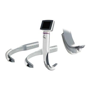 King Vision Video Laryngoscopes are affordable, durable and portable. They are ideal for indirect laryngoscopy, difficult endotracheal intubations as well as routine intubations. The King Vision accommodates minimum mouth openings of 13mm for the standard blade and 18mm for the channeled blade making it effective for the majority of adult patient populations. Features: Ergonomics, durability and technique of a traditional rigid laryngoscope Affordability never seen in the market before High-quality imaging of FOBs and other indirect laryngoscopy equipment with an attached, reusable, OLED video display for exceptional color, contrast and brightness Portability of hand-held video laryngoscopes Convenience and sanitation of disposable, channeled and non-channeled blades that incorporate a camera and light source — no need for a stylet with channeled blades Clarity and accuracy of a LED light and CMOS camera Technical Specifications: Batteries: 3AAA Battery Life: <90 min. (confirm with Power Indicator status) Computerized Power Management System: Auto Shut off, Auto white balancing Video Aspect Ratio: 4:3 Video Port: RCA connection to monitor with cable Video Refresh Rate: 30 frames per second Video Resolution: 320 x 240 (QVGA) pixels per frame Video Screen: OLED Display Video Screen Siz:e 6.096cm / 2.4″ diagonal Viewable Angle: 160° Anti-Fog: Coating on distal window Maximum AP Height of Blade: <13mm / .51″ (non-channeled); <18mm / .7087″ (channeled) Blade Width (at mouth): 26mm / 1.0236″ (standard), 29 mm / 1.1417″ (channeled), 16 mm / .6299 at distal tip Camera Chip: CMOS Camera Resolution: 640 x 480 VGA Disposable Material: Polycarbonate / ABS, anti-reflective, coating on display window ET Size; 6.0mm – 8.0mm / .2362″ – .3150″ Light Source: White LED Maximum Pull Force on Blade: 15lbs Click Here To Download Catalogue
King Vision Video Laryngoscope
-
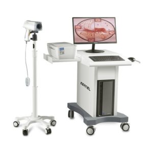 Video Colposcope is a gynecological clinical diagnostic instrument. For the diagnosis of cervical disease, it can be observed 10 to 60 times magnification images, found small lesions the naked eye can not find. Through this amplification effect, the doctor can clearly see the cervix blood vessels on the skin, detection of cervical cancer precursor lesions, Features : 1. Camera 1/4 inch CCD, 800,000 pixels 18x optical magnification and 12x digital magnification Horizontal resolution: 700 line Focus: manual or auto. 2. Control Handle Humanized operation panel, can realize zoom, focus, imaging mode, image marking, brightness adjustment and other functions with single hand. 3.Camera holder 360°adjustable full-damping camera holder, can be automatically fixed at any Angle. Software: English, Spanish, Russian, Vietnamese, Italian, Turkish, French. Specifications: Lens lighter type: LED Lens video-output: PAL, VBS-standard 1.0Vp-p Power Requirements: AC220V ±10% Power Rating: 500W Brightness adjustment: Automatic electronic shutter Ambient temperature: -40°C to +50°C Storage temperature: -20°C to +60°C Relative Humidity: 30~80%RH Configuration: SD Camera software + Vertical stand PC: 516usd 2.5 inch screen Assemble Trolley Accessories
KN-2200 Video Colposcope
-
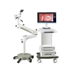 Colposcope is a gynecological clinical diagnostic instruments. For the diagnosis of cervical disease, it can be observed 10 to 60 times magnification images, found small lesions the naked eye can not find. Through this amplification effect, the doctor can clearly see the cervix blood vessels on the skin, detection of cervical cancer precursor lesions, Feature: High eyespots, wide field eyepieces, eyepiece has two choices 0 degrees and 45 degrees for different height of bed examination, make the clinical operation more comfortable. Up to 30 times optical zoom, relying on high-quality amplification effect.It can be clearly observed diseased various small cells , can carry on the accurate positioning biopsy for pathological changes of living, help to improve the judgment before all kinds of cervical disease, cancer screening accuracy, reduce the misdiagnosis. Photoelectric and push the overall integration design, flexible operation, reasonable layout, small footprint, in limited space can meet the clinical application. Technical level of all the joints to adopt damping bearing, lifting arm can make the different height of users easily adjusted, unique freedom arm technology can make the microscope accurate and stable, lifting, spatial arbitrary point fixation. Specifications: Binocular: 12.5x, 45degree; Optional: 12.5x, 0 degree;20x, 45 degree; 20x,0 degree Focal distance: 45 degree binocular: 125mm; 0 degree: 160mm The Minimum magnification is 17x, and Max Magnification is 32x. depend on different binocular. Configuration: ⚫ Optical and Image System ⚫ Swing Arm ⚫ Cold LED Light Source ⚫ CCD+ Software (With foot-switch and Built-in Capturing Card) ⚫ Computer and LCD displayer ⚫ Printer ⚫ ABS Trolley
KN-2200B (LED light) Colposcope
-
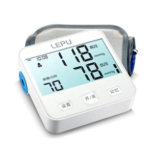 Features 2200mAh lithium battery Monitor algorithm, for more accurate measurement 5.2-inch large backlit screen, making health parameters clearly visible Cuff recognition function, reducing the measurement error
Lepu 70D Sphygmomanometer
Zenrox Healthcare Solutions - Advanced Medical Equipment in Lagos
Zenrox offers cutting-edge medical equipment and comprehensive support services in Lagos. Explore our products designed for superior healthcare delivery. Contact us today for innovative medical solutions.