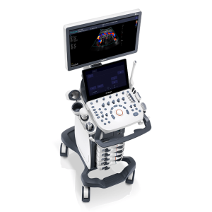
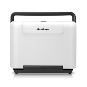
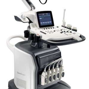
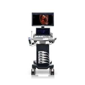
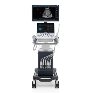
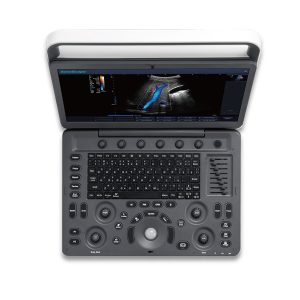
Sonoscape E3 Ultrasound Machine

Categories: , , SKU: ZN2024330 Tags: ,

6,100,000.00

A brand new portable ultrasound system E3, brings you a distinct experience with SonoScape’s traditional imaging technologies. E3’s accurate B mode and sensitive color signal give crisp, detailed images to improve your scanning experience while increasing your diagnostic confidence.

Features and Benefits

C-field Beam: The continuously dynamic focus provides more energy which contributes to higher contrast resolution, signal-noise ratio and uniformity in the image.
μ-Scan: The latest generation of μ-Scan imaging greatly enhances the image by reducing noise, improving signal strength and improving visualization.
SR Flow: With SR Flow the sonographers can easily see in detail very small veins and slower velocities for the detailed blood flow information of the patient.
Tissue Specific Imaging: The system detects different tissues automatically by matching different acoustic ranges, from which the user can then acquire images with more uniformity and higher spatial resolution.

15.6″ Anti-flickering HD LED Screen
Color Doppler system
Standard two-active probe sockets

Materials Included

Ultrasound system with LED Screen
Backlit Keyboard and Intelligent Panel
3 Transducer ports
Long-lasting Battery

Technical Specifications

Design

15.6″ LED Monitor
Two Probe Socket

Display Modes

B, 2B and 4B
M and B/M
Pulsed Doppler
Tissue Harmonic Imaging
DPI
ECG
Trapezoidal
µ-scan: 2D speckle reduction
M and B/M dual life

Probes

Micro-convex C611 4-8 MHz
Linear L741 5-15 MHz
Low Frequency phased array 2P1 2-4 MHz
High Frequency phased array 5P1 4-7MHz
Rectal Probe L741 5-15MHz

Settings

Zoom: up to 4x
2 line-density levels
TGC slider controls: 8 levels
20 – 255 dB dynamic range
Focus span adjustable: 5 levels
Persistence: 8 levels
Chroma: 13 levels
Scan depth: up to 26 cm

Click Here To Download Catalogue

Description

  • C-field Beam: The continuously dynamic focus provides more energy which contributes to higher contrast resolution, signal-noise ratio and uniformity in the image.
  • μ-Scan: The latest generation of μ-Scan imaging greatly enhances the image by reducing noise, improving signal strength and improving visualization.
  • SR Flow: With SR Flow the sonographers can easily see in detail very small veins and slower velocities for the detailed blood flow information of the patient.
  • Tissue Specific Imaging: The system detects different tissues automatically by matching different acoustic ranges, from which the user can then acquire images with more uniformity and higher spatial resolution.
  • 0/5 (0 Reviews)
    0/5 (0 Reviews)