Only logged in customers who have purchased this product may leave a review.
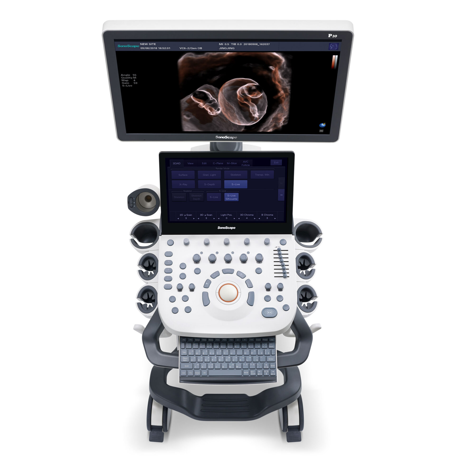
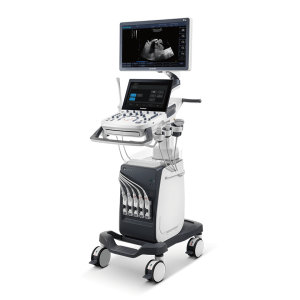
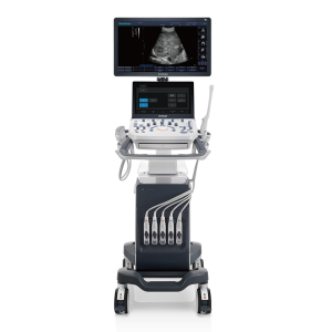
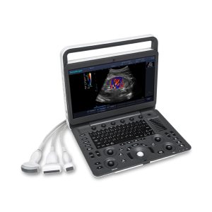
Sonoscape P20 Ultrasound Machine
Available in stock
Reviews (0)
Zenrox Healthcare Solutions - Advanced Medical Equipment in Lagos
Zenrox offers cutting-edge medical equipment and comprehensive support services in Lagos. Explore our products designed for superior healthcare delivery. Contact us today for innovative medical solutions.
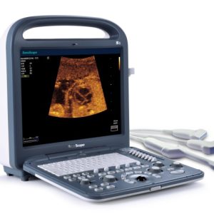
Available in stock
Only logged in customers who have purchased this product may leave a review.
Reviews
There are no reviews yet.