-
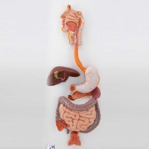 Life-size human digestive system model that demonstrates the entire digestive system in graphic relief. Digestive system features: Nose Mouth cavity and Pharynx Esophagus GI tract Liver with gall bladder Pancreas Spleen The duodenum, caecum and rectum of the digestive system are opened. The transverse colon and front stomach wall are removable from the digestive system for detailed study of the anatomy. This high quality digestive system is a great teaching tool for any anatomy lesson or doctor’s office. Digestive system mounted on baseboard for easy display in the classroom.
3B Scientific Human Digestive System Model, 3 part
-
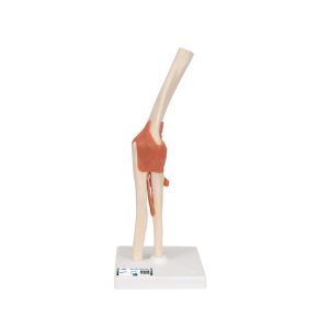 This high quality, life-size functional elbow joint model provides a graphic demonstration of the anatomy and mechanics of the human elbow joint. Use this fully flexible model to demonstrate abduction, anteversion, retroversion and internal/external rotation. Elbow joint consists of portion of the humerus, complete ulna and radius as well as joint ligaments. Comes on removable stand for easy study or display.
3B Scientific Functional Human Elbow Joint Model with Ligaments – 3B Smart Anatomy
-
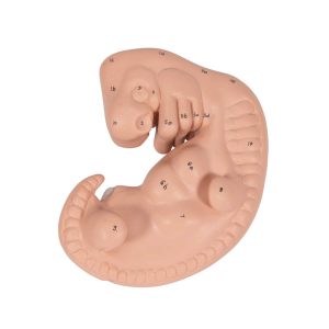 This human embryo model shows the anatomy of an embryo at approximately 4 weeks old. At 25 times life size this human embryo is great for studying human development. The high quality model is affordable without sacrificing any anatomical detail.
3B Scientific Human Embryo Model, 25 times Life-Size – 3B Smart Anatomy
-
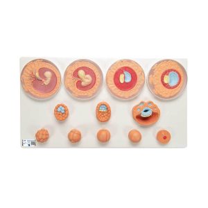 The model represents the development of the human germ cells from fertilisation until the end of the 2nd month of pregnancy in 12 stages. Each stage can be removed from the common stand as an individual part and can be purposefully used for teaching and tests for the embryological specialist field. Ovum at time of fertilisation (conception) with male gamete (sperm) Zygote at 2-cell stage, approx. 30 hours after fertilisation Zygote at 4-cell stage, after around 40-50 hours Zygote at 8-cell stage, after around 55 hours Morula Blastocyst after around 4 days Blastocyst after around 5 days Blastocyst after around 8-9 days Germ cells at approx. 11th day Germ cells at approx. 20th day Embryo at around the end of the 1st month of pregnancy Embryo at around the end of the 2nd month of pregnancy
3B Scientific Embryonic Development Model in 12 Stages – 3B Smart Anatomy
-
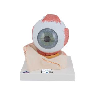 Removable parts of this anatomical human eye model include: Upper half of the sclera with cornea and eye muscle attachments Both halves of choroid with iris and retina Lens Vitreous humour This eye model is great for studying the anatomy of the human eye! Eye on base of bony orbit.
3B Scientific Human Eye Model, 5 times Full-Size, 7 part
-
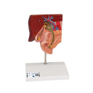 This graphic gallstone model for patient education shows the anatomy of the biliary system and its surroundings in half natural size. Both acute inflammation (cholecystitis) and the tissue changes caused by chronic inflammation can be identified in the gallbladder wall. Gallstones can be found in the following typical locations: In the fundus area of the gall bladder In the area of the spiral valve In the area of the common bile duct In the papillary opening to the small intestine Gallstone model mounted on base.
3B Scientific Gallstone Model – 3B Smart Anatomy
-
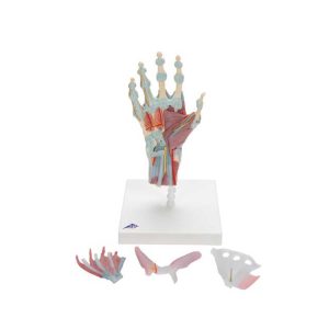 The bones, muscles, tendons, ligaments, nerves, arteries, and veins are all featured in this high quality 4 part model of the hand and lower forearm. The dorsal side of the hand shows the extensor muscles as well as portions of the tendons at the wrist as they pass under the extensor retunaculum. The palmar face of the hand is represented in three layers, the first two are removable to allow detailed study of the deeper anatomical layer of the hand. In addition clinically important structures such as the median nerve and superficial palmar arterial arch can be explored in detail in the hand model. The deepest anatomical layer allows for study of the intrinsic muscles and deep palmar arterial arch in addition to other details of the anatomy of the hand. This high quality anatomically correct hand model with ligaments and muscles is great for detailed study.
3B Scientific Hand Skeleton Model with Ligaments and Muscles – 3B Smart Anatomy
-
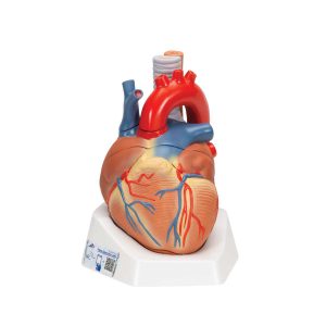 This heart model shows the anatomy of the heart model and is horizontally sectioned at the level of the valve plane. The following parts can be removed from the heart: Esophagus Trachea Superior vena cava Aorta Front heart wall Upper half of the heart This high quality model clearly shows over 30 different anatomical regions in the heart. Comes the product manual for easy identification of anatomical features.
3B Scientific Human Heart Model, 7 Part – 3B Smart Anatomy
-
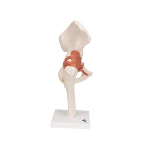 This life size functional hip joint model clearly shows the anatomy and mechanics of the human hip joint. This fully flexible hip joint demonstrates abduction, anteversion, retroversion and internal/external rotation. This high quality functional joint consists of a portion of femur, hip bone and joint ligaments. Comes on a stand for easy display in the classroom or doctor’s office.
3B Scientific Functional Human Hip Joint Model – 3B Smart Anatomy
-
 The MICROanatomy™ Eye model illustrates the microscopic anatomical structure of the retina with choroid and sclera. The left block-like, layered side of the eye model shows the complete structure of the retina including the supplying vascular layer and parts of the sclera from a light microscopic view. The right part of the eye model is a sectional enlargement. MICROanatomy™ Eye shows the microscopic structure of the photoreceptors and the cells of the pigmented layer. Left part of MICROanatomy™ Eye 850-times enlarged – right part 3800-times enlarged. You’ve never seen the human eye like this before!
3B Scientific 3B MICROanatomy™ Human Eye Model – 3B Smart Anatomy
-
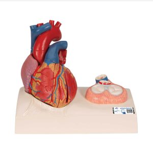 This model is cast from a real human heart and didactically prepared to facilitate a better understanding of the anatomy and blood flow of the heart. It shows the cardiac valves during diastole and on the base the valves are shown in systole. A dissection through the median plane makes an easy demonstration possible. Its attention to detail and high quality craftsmanship makes it definitely the top of the line heart model. The following feature set makes it stand out from the crowd and a must have for any health professional. All of the original heart structures were successfully obtained during the time consuming and detailed casting procedure making this model highly accurate and lifelike 2 atria and 2 ventricles show all the normal anatomical structures of the papillary muscles and heart valves Uniquely dissected in the median plane to optimally demonstrate the path of the oxygenated and deoxygenated blood The heart model shows both the diastolic and systolic state. In the model itself the valves are shown in the diastolic state and in the detail view on the base the valves are shown in the systolic state The heart valves are made of elasticated plastic making them very durable The base displays the heart in its natural position in the human body Life size cast from real human heart Easy and fun to use magnetic assembly (5 pieces) for easy demonstrations
3B Scientific Life-Size Human Heart Model, 5 Parts with Representation of Systole – 3B Smart Anatomy
-
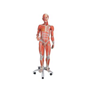 This muscle figure is the finest teaching tool available! Standing over 4 1/2 feet tall, this 3/4 life-size human replica depicts deep and superficial musculature in addition to the body’s major nerves, vessels, tissues and organs in exquisite detail. The internal organs are removable (45 pieces in all) to reveal the fundamental interrelationships of human morphology. Remove the calvarium to view the 3-part removable brain. Look beneath the liver to reveal the gallbladder and bile duct. Peer inside the appendix, stomach lungs, heart or kidney. Remove and view the details of 13 different muscles of the arms and legs. This dual sex muscle figure version has interchangeable genital inserts and a female mammary gland as well as a detailed multilingual product manual identifying over 600 hand-numbered structures. Hand-painted and mounted on a convenient roller base. Includes the following features: 5 arm/shoulder muscles 8 leg/hip muscles 2-part removable heart 5-part head with removable brain 2-part removable lungs 2-part stomach Removable 4-part male and 2-part female genital inserts Detachable arms, leg, head, and abdominal wall for detailed study Now on a stable metal stand with 5 casters!
3B Scientific 3/4 Life-Size Dual Sex Human Muscle Model on Metal Stand, 45-Part – 3B Smart Anatomy
-
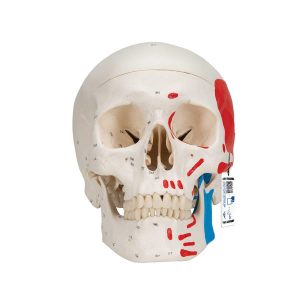 This replica of the human skull is a fantastic tool for teaching and learning the anatomy of the skull. The muscle origins (red) and insertions (blue) are shown in color on the left side of the skull. Cranial bones and structures are numbered on the right side. Jaw hinges with spring to simulate real movement. This skull model identifies over 140 anatomical details. High–quality original casts Skull is handmade of hard, unbreakable plastic Highly accurate representation of the fissures, foramina, processes, sutures etc. Can be disassembled into Skull Cap, Base of Skull and Mandible As an option, you can insert a 5–part brain (C 18) into the skull Now with magnetic connections This high quality skull is a great anatomy teaching tool!
3B Scientific Classic Human Skull Model painted, 3 part – 3B Smart Anatomy
-
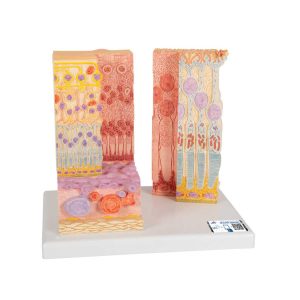
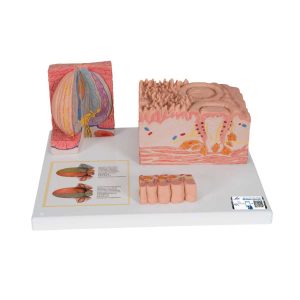 The latest model in our 3B MICROanatomy™ series, the tongue, is fascinating in that it combines various enlargements of specific parts of the tongue in one model. It comprises a macroscopic view of the tongue in life size (dorsal view) and microscopic views of the various papillae of the tongue (10-20x life size) and of a taste bud (approx. 450x life size). All views are mounted on a base that also features an overview of the sensory and sensitive innervation of the tongue. A unique model for an intensive study of the tongue.
3B Scientific 3B MICROanatomy™ Human Tongue Model – 3B Smart Anatomy
-
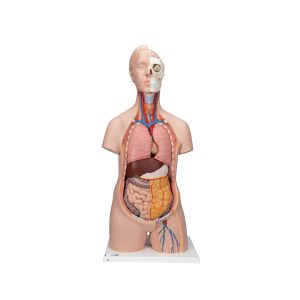 This 12 part anatomically correct human torso is an educational tool of true quality. The unisex torso is hand–painted true to detail and made of high–quality plastic. This classic human torso was developed and modeled in Germany. Whether you are a student studying human anatomy in a biology classroom or a doctor explaining something to a patient, this human torso model is a valuable tool. The following components of this unisex torso are removable: 2–part head 2–part removable heart 2 lungs Stomach Liver with gall bladder 2–part intestinal tract Front half of kidney All the organs in this human torso are hand painted for a quality product. This great human anatomy educational tool and makes learning the location of the human organs easy. It is also supplied with the 3B Torso Guide.
3B Scientific Classic Unisex Human Torso Model, 12 part – 3B Smart Anatomy
-
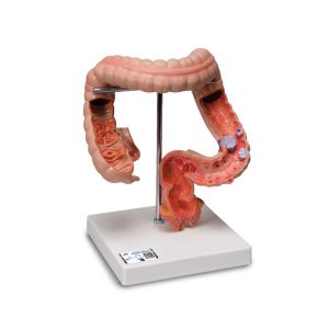 This model shows the usual benign and malignant changes in the colon and rectum. These include diverticula, polyps, hemorrhoids, fistulae, fissures, chronic inflammatory diseases (Crohn’s disease and ulcerative colitis), as well as intestinal cancer in various stages.
3B Scientific Intestinal Diseases Model
Zenrox Healthcare Solutions - Advanced Medical Equipment in Lagos
Zenrox offers cutting-edge medical equipment and comprehensive support services in Lagos. Explore our products designed for superior healthcare delivery. Contact us today for innovative medical solutions.