-
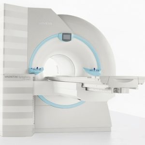 More power toyou. MAGNETOM® Symphony® and Tim (Total imaging matrix) technology -a perfect combination or patient-friendliness and fast scanning capabilities. 5T, 60-cm MR system incorporating Tim technology Available in an 18- or 32-channelcongregation Expands upon the Symphony applications, adding additional inline processing such as Inline Diffusion or automated ADC mapping Nine Tim application suitesstandard, including cardiac,pediatric, and breast Fully integrated coildesign Lightweight (patient-friendly), scalable coils part of the standard congregation
MAGNETOM®
-
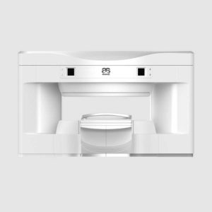 OPENMARK 5000 is 0.51T MRI. It’s approved by FDA and has CE mark. It adopts two-pillar magnet design with 280 degree openness and equipped with powerful RF and gradient system, together with advanced imaging technology, making it as a high-end system which is comparable to high-field MRI. Features: With the highest system stability and the highest homogeneity of the magnet field in permanent MRI Screens on both sides facilitate positioning; 280 degree two-pillar magnet design ensures stable magnet structure and facilitates interventional treatment. Active and passive shimming calibrate technology ensures the magnetic field uniformity Motor-driven patient couch makes it easier for patients to access and for positioning Powerful hardware and software platforms ensure the scan speed, image quality and make it possible for advanced imaging functions Fast scan speed eliminates motion artifact Rich scan sequences, advanced imaging technology and powerful postprocessing technology ensure image quality, extend more applications, which can fully satisfy the clinical needs Intelligent user-friendly operating system ensures you easy operation Technical Specifications: No. Technique Parameter 1 Magnet System 1.1 Magnet Type Permanent Magnet Automatic constant temperature system 1.2 Field Strength 0.51T 1.3 Magnet Shape Dual-pillar shape 1.4 Homogeneity(40cm,DSV,VRMS) ≤1.6ppm 1.5 Shim Method Active/Passive 1.6 Magnet Vertical Gap (Cover) 40cm 1.7 Magnetic Pole Dia. (Exclude Cover) 145cm 1.8 Accessibility(Horizontal Opening Angle, 280° 1.9 5 Gauss fringe field X-axis:horizontal ≤2.5m Y-axis:Vertical ≤2.5m Z-axis:horizontal ≤2.5m 2 Patient Couch and Communication 2.1 Patient Couch Driven mode Motor-driven 2.2 Max. Patient Weight ≥200kg(440lbs) 2.3 Patient Positioning Tools Laser Light Localizer for positioning of center slice Motor-driven transfer to center of imaging volume 2.4 Position accuracy ±1mm 2.5 Emergency Call Key Yes 2.6 Intercom System Yes 3 Gradient System 3.1 Gradient Field Strength(Single Axis) ≥30mT/m 3.2 Gradient Slew Rate (Single Axis) ≥100mT/m/ms 3.3 Rise Time ≤0.3ms 3.4 Gradient Cooling System ( Gradient coils and Power electronics) Air Cooling 4 RF System 4.1 RF System Type Digital Transmit and Receive signal 4.2 Number of RF Channels 4 4.3 Transmitter Amplifier Peak Power 6kW 4.4 RF Bandwidth of Receiver ≥1.25MHz 4.5 Head Coil Yes 4.6 Neck Coil Yes 4.7 Body/Spine Coil (17 inch) Yes 4.8 Body/Spine Coil (21 inch) Yes 4.9 Knee Coil Yes 4.10 Shoulder Coil Yes 4.11 Flexible Coil Optional 4.12 Breast Coil Optional 5 Computer System 5.1 Host Computer DELL Computer (for MR) 5.2 System Software Windows XP 5.3 Operation Software APEX 5.4 CPU Clock rate 3.0GHz 5.5 Main Memory 4GB 5.6 Color LCD Monitor 19” 5.7 Keyboard and Mouse Standard 5.8 Image Reconstruction Speed(256 x 256 Matrix) 200 frame/Sec. 5.9 Hard Disk 500GB 5.10 Image Storage Capacity(256 x 256 Matrix) 500,000 5.11 Media Driver DVD RW 5.12 DICOM 3.0 Yes 5.13 Ethernet Yes 5.14 Operation Console Yes 5.15 Operation Chair Yes 6 Scanning Parameter 6.1 Max. FOV 410mm 6.2 Min. FOV 5mm 6.3 Min. TE(SE) 5ms 6.4 Min. TR(SE) 11ms 6.5 Min. TE(GR) 1ms 6.6 Min. TR(GR) 3ms 6.7 Min. 2D Thickness 1.0mm 6.8 Min. 3D Thickness 0.1mm 6.9 Max. Image Matrix 512×512 7 Scanning Sequence & Imaging Technique 7.1 Spin Echo 2D/3D (SE 2D/3D) Yes 7.2 DE/QE Yes 7.3 Fast Spin Echo 2D/3D(FSE 2D/3D) Yes 7.4 Single Shot FSE 2D/3D Yes 7.5 Inversion Recovery(IR) Yes 7.6 Fast Inversion Recovery(FIR) Yes 7.7 Gradient Echo 2D/3D(GR 2D/3D) Yes 7.8 Fast GR 2D/3D Yes 7.9 SPGR Yes 7.10 FLAIR Yes 7.11 Fat Imaging Yes 7.12 Fat Suppression imaging Yes 7.13 Water-Fat Separation imaging Yes 7.14 TOF MRA(2D/3D) Yes 7.15 MRCP(2D/3D) Yes 7.16 MRU (2D/3D) Yes 7.17 MRM Yes 7.18 Fast Hydrograph Imaging Yes 7.19 Diffusion Weighted Imaging(DWI) Yes 7.20 Max. b Value 1000s/mm2 7.21 Breath Hold Technology Yes 7.22 Magnetization Transfer Contrast(MTC) Yes 7.23 Multi-slice and Angle-free Presaturation Yes 7.24 Saturation Tracking Yes 7.25 Maximum Intensity Projection(MIP) Yes 7.26 Multi-Angle Projection(MAP) Yes 7.27 3D Reconstruction Yes 7.28 Multi-planar Reconstruction(MPR) Yes 7.29 Multi-Artifacts Eliminating technology Yes 7.30 Checking with Part Metal Implant Yes 7.31 Online Image Filtration Yes 7.32 Online Post Procession Yes 7.33 3D Scout Yes 7.34 Scanning Protocol Preset Yes 7.35 Scanning Protocol Queue Waiting Yes 7.36 Advanced Image Post Processing Yes 7.37 Image Fusion Technology of Vascular Yes 7.38 Image Fusion Technology of Spine Yes Click Here To Download Catalogue
Anke MRI Openmark 5000 Permanent System
-
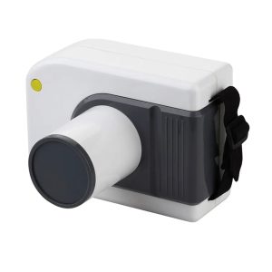 Features: 1. The device is DC high frequency portable dental unit, small, light and nearly no radiation. 2. There are manual buttons installed on the surface of shell, operate easily, all of components are installed in the central PC board, insulation vacuum and sealed stereotype protection make its brilliant features. 3. The unit is mainly suitable for oral pre-treatment diagnosis of internal organisation structure and root depth and so on. It is indispensable in clinics especially for implant surgery. 4. The battery is durable, it can take about 5 hundred images after fully charged. 5. It can be connected with intra-oral digital X-ray imaging system. 6. 3 Colors Specifications: Tube Voltage 60kV Tube current 1MA Exposure Time 0.02~2S Tube Focus 0.3*0.3mm Frequency 30kHz Battery DC14.8V 6400mA Rated Power 60VA Charger Input AC100V-240V± 10% Charger Output DC16.8V Gross weight 3.5KG Package size 220*240*275 The distance from skin to cone 130mm
Portable X-Ray Unit
-
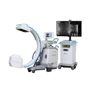
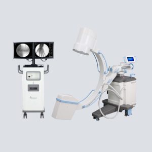
 Our Neeo “C” arms are easy to place, use and are specifically designed to be used in orthopedics, traumatology, abdominal surgery, urology, cardiology and operating rooms. Using Neeo with the RTP (Real Time Processing) option it is possible to perform vascular, urological and cardiological diagnostics. One of the main functions, digital image subtraction, allows to see, as an example, the passage of contrast liquids in a tissue or in a venous or arterial duct; thanks to the possibility of looping, the acquired video can be reproduced several times to monitor more accurately the passage of the fluid within the area in question. Angiographic measurement is another useful function in the vascular field (QA Quantitative Angiography) that allows the measurement of stenoses. Finally, fluoroscopy allows the correct positioning of stents or expanders. Neeo is used in various interventional and diagnostic procedures in traumatology and orthopedics wards and operating rooms as well. Thanks to low-dose fluoroscopy, it is possible to use the device for positioning bone or subcutaneous grafts, inserting K-wire (Kirschner wire) for stabilization of bone fragments or for the correct positioning of prostheses. The low dose emitted ensures safe use for both the patient and the surgeon or doctor on the operating field. On the control panel there is a large touch screen display that allows to adjust the basic functions of the equipment. From this display it is possible to select and adjust the fluoroscopic data for the examination, activate or deactivate the laser pointer, select between pulsed, one shot or standard fluoroscopy, rotate the image and perform all operations on collimator. The four side buttons on the display offer the possibility to move the bow vertically thanks to an extremely silent motor. Neeo has two 19 “medical grade monitors that can be positioned according to the needs of the medical practitioner. Work monitors and feedback monitors are separated to be managed independently. The possible movements are: rotation, revolution, tilting and possibility of height adjustment. Features: Continuous fluoroscopy Pulsed fluorography One Shot fluoroscopy Low dose fluoroscopy Radiography with cassette (up to 5KW) 110,000 images of memory (Standard) 19 ″ Medical Flat Monitor (X2) Touch control panel Image Processing – 1K X 1K, 16 bit DAP-Dose Area Meter DSA Road Mapping Laser Marker Technical Specification: Power: 5KW kV Range (kV): 40 – 120 mAs Range (mAs): 1 – 250 SID (mm): 1000 Free Space (mm): 730 DICOM Feature: RTP-SW DSA RTP-SW QA RTP-SW Worklist RTP-SW Print RTP-SW CD DVD Saving RTP-SW MPPS RTP-SW Query/Retrieve RTP-SW HCF RTP-SW BASE RTP-SW STCOMMIT Configuration: C-Arm Structure, Trolley for Monitor, Collimator, Monoblock, Toshiba 12” Image Intensifier, 19″ Philips Medical Flat Monitor (X2), Camera CD1030CA 1KX1K, Sony Printer UP-X898MD, Cassette Holder Click Here To Download Catalogue
IBIS Neeo R9 Digital Surgical C-Arm
-
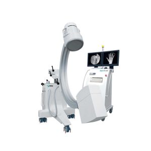
 The OSCAR 15 is a culmination of several years worth of developmental experience from Genoray. With CMOS imaging excellence & 15kW HFG you diagnosis need will be met while improving your productivity especially DSA (Digital Subtraction Angiography). APPLICATION General Surgery Office based Vascular Center Pain Management Orthopdics Urology Cardiac Procedures Hybrid OR Neuro & Spine Surgery Pain Management Orthopedic Surgery Trauma Procedure -Urology Procedure Cardiac Surgery Peripheral Artery Diseases -Vascular Surgery SPECIFICATION 260 x 260 mm CMOS Type Flat-Panel detector for distortion-free imaging (High resolution images, Wide FOV, Low Noise) 15kW HFG 4″Touch LCD monitor 43″ LCD Monitor Dual Foot Switch DICOM 3.0 -CD/DVD Burner – USB Port 800mm free space and 150° (+90°,-60°) orbital rotation, SID 1,000mm 155º Dynamic Orbital Rotation 2 kW Stationary Anode X-Ray Tube 5 Million Images Storage Capacity FEATURE Exceptional Image Quality With an optimal flat panel detector size of 26 x 26cm you won’t miss a thing with its high quality resolution. It makes accurate diagnosis in a variety of departments especially DSA Low Dose Mode Low dose mode is desgined to acquire reasonable image to diagnose the patient with minimum dosage. Edge Enhancenment For user to get more accurate diagnosis result enhancing edge of image. Motion Correction This function detects the movement and reduce the after image while exposing X-ray. Metal Correction To prevent over-dose radiation or low quality image casued by metal inturrption on field of view. Virtual Collimator The virtual collimator allows for the selection of your desired field of view, while reducing the amount of radiation exposure by limiting the X-Ray beam. Auto Collimation Prevention of unncessary X-Ray exposure by focusing on the area of interest while autmoatically collimating the remaining areas. POWERFULL SOFTWARE ZENIS A total solution from acquisition, storage, management, communication to print out. Provide convenient environment from user-centric interface. Diagnosis and confirm from recognizable simple icons. Convenience of database management. Convenient diagnostic functions for easy patient / image management Accurate diagnostic tools Improve the efficiency of your hospital management Perfect compatibility with all PACS A must have for a digitally equipped hospital Convenient communication and management for your customers Dicom Support DIGITAL SUBTRACTION ANGIOGRAPHY Native DSA Pairing fluoroscopy with constrast media to display the basic angiography views Motion Matching Selects the proper mask to apply and remove artifacts made by a patient’s movement or breathing Post-Processing Processing: Improvement of the processed image after the DSA procedure Landmarking / Brightness / Contrast After setting the position for a vessel, the subject can be placed back to their original position by using the shift function to compensate for any movement. Allows for various functions that assist with accurately inserting a catheter. Peak Opacification Ability to diagnose a blood vessel with only a small amount of contrast media Road Mapping, Land Mark After setting a position for a vessel, the subject can be moved back to their original place by using the shift function to compensate for any movement. Provides various functions that helps accurately to insert a guide wire, catheter is compatible with the hybrid operating room. Auto Roadmap Mask Obtain blood vessel type information while only using a small amount of contrast media Manual Roadmap Mask Roadmap your vessels using a prevoiusly taken DSA image Roadmap Pixel Shift Re-position the roadmap mask by shifting the pixels to the proper position Click Here To Download Catalogue
Genoray OSCAR 15 Surgical C-Arm Machine
-
 Fluoroscopy Powerful, Light and Versatile Performance with the 9″ high-resolution image intensifier and million pixel (1K x 1K) high-definition camera, the Zen-7000 supports clear images without distortion to provide the best diagnostic imaging environment possible. Exceptional Image Quality 9″ or 12” image intensifier 1K x 1K High resolution CCD Camera Wide FOV (Field-of-View) Low Dose, Low Noise High-Definition High Contrast Rotating Anode Tube X-Ray Dose Management Low Dose Mode Auto / Virtual Collimator Iris Collimator Compact & Intelligent Design Lightweight Architecture Intelligent operation Compact, Hybrid Body 7″ Touch Screen LCD Monitor 19-inch LCD Two Monitor Powerful Software Image Management DICOM 3.0 Database Management Approved Format Air Cooling High Performance Protection from Overheating and Damage Click Here To Download Catalogue
Genoray ZEN-7000 Surgical Mobile C-Arm
-
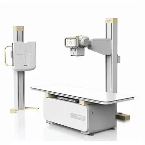 Product Details The GXR diagnostic X-ray system matches with a radiographic room which perfectly fits your workflow. It can be easily upgraded to a DR system with the help of the DR interface and PC interface in the GXR generator, as well as the FPD(Flat Panel Detector) compatible Bucky. The GXR X-ray system is equipped with a high frequency X-ray generator which consistently produces high quality radiographs in favor of high quality X-ray output, with a very small kV ripple and accurate mA & mAs. The GXR X-ray system is designed to provide convenience to the operator and comfort to the patient. OPTIONS AEC DAP(Dose Area Product) Meter Vertical tube synchronization Table Bucky auto tracking Lateral cassette holder WBS external cassette holder Line Laser Handgrips (Tabletop, WBS – Chest, Overhead) Electromagnetic switch for Tube Stand Rotation (FC or FM) 23 inch Touch Screen Console
DRGEM GXR Diagnostic X-ray System
-
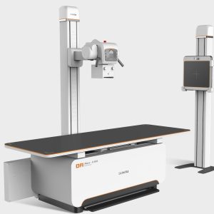
Feature
 Innovative User-friendly Design
- 360°rotation of the X-ray tube column for any special radiographic positions.
- Pluggable detector and grid design.
Functional Applications- Automatic Exposure Control(AEC) function(Optional).
- Standard Dose Area Product(DAP) function.
- Pneumoconiosis Anatomic Procedural Radiography (APR) provides chest X-ray screening for pneumoconiosis.

 Stable Input Power Guarantee
- SONTU Power Solutions break limited input power with a single-phase option available (Optional). Excellent performance under various electrical and installation circumstances.
Multiple Configurations- Providing 50kW and 63kW high-frequency generator options.
- 14*17/17*17, wired/wireless flat panel detectors available.

Clinical Image
Sontu Statics X Ray sontu-100rad RAD(S) Series Floor-mounted DR
Zenrox Healthcare Solutions - Advanced Medical Equipment in Lagos
Zenrox offers cutting-edge medical equipment and comprehensive support services in Lagos. Explore our products designed for superior healthcare delivery. Contact us today for innovative medical solutions.