-
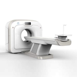
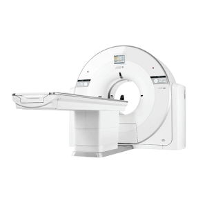 Technologies Ultra-Low-Noise Z-Detector The ultra-low noise detector fundamentally improves the image quality. With Through-Silicon-Via (TSV), Z-Detector enables direct output of digital signal, which achieves not only overall noise reduction, but also helps to ensure optimal image quality with low radiation dose. Iterative Denoising Technique The customizable KARL 3D iterative denoising algorithm maintains consistent image quality with a reduced dose. KARL 3D can also be combined with the metal artifact correction (MAC) algorithm to process scan data of patients with metal implants, eliminating metal artifacts and reducing image noise. Dedication to Fine Craftsmanship and Quality United Imaging has a remarkable dedication to craftsmanship and quality. From every detail inside the scanner to the finished product, the uCT 530 exudes fine craftsmanship and quality, which is evident in the stunning clinical images produced by the system. Highly Efficient Workflow High Throughput Hardware Platform The uCT 530 is equipped with a 5.3 MHU X-ray tube with fast heat dissipation capacity of up to 815 kHU/min, which ensures a fluid clinical workflow and offers high throughput. Easy-Logic Intelligent Prediction Platform The Easy-Logic intelligent prediction platform can predict the user’s next operation and initiate system preparation ahead of time. Factors such as tube preparation and early rotation of the gantry enable a seamless and efficient scanning process. uECO Energy-Saving Module The uECO shortens the time needed to stabilize the system after standby. The energy-saving module reduces the power consumption of the CT and HVAC systems. Poised. Proficient. Clinical Gallery Abdomen & Pelvis Study, Coronal View Carotid and Circle of Willis Study Brain uCT 530 BRAIN ABDOMEN & PELVIS STUDY, CORONAL VIEW CAROTID AND CIRCLE OF WILLIS STUDY BRAIN ABDOMEN & PELVIS STUDY, CORONAL VIEW CAROTID AND CIRCLE OF WILLIS STUDY BRAIN Passion For Change
United Imaging CT Scan UCT 530
-
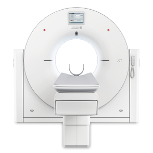 Technologies Z-Detector The Z-Detector reduces electronic noise down to a single photon level and the fully integrated design shortens the signal transmission length from centimeter to micron scale, resulting in tremendous electronic noise reduction and efficient X-ray utilization. KARL 3D Iterative Denoising The algorithm is performed in both the projection and image spaces to reduce dose while preserving image quality. Workflow with Ease EasyRegister The most frequently used protocols are placed in the front page shortcut area for easy selection. Along with the preset patient positions, the patient registration can be completed efficiently. EasyScan Intelligently plans the exam for individual patients. Parameters such as the scan range, reconstruction FOV and gantry tilt angle are automatically adjusted.* * Optional EasyLogic The system anticipates the operator’s needs and prepares itself to reduce delays. Minimizes the wait time between operation procedures and accelerates CT exam workflow significantly. Imaging with care 70kV Scan Mode 70kV Dose reduction can increase SNR for Iodine-based contrast agents especially for small BMI or younger patients. uDose 3D Dose Modulation Based on anatomic structure information, it generates optimized 3D dose distribution plan. Outstanding Financial Performance Reducing total cost of ownership for operation. Reliable Components Light-weight tube with reliable design and good serviceability. Self-cleaning carbon brushes for improved slip-ring reliability. Go Green Switching the system into uECO mode during night or when the system is idle saves 30% power compared to conventional standby modes. Small Footprint uCT 528 requires a minimum installation area of 9m2. Clinical Gallery uCT 528
United Imaging CT Scan UCT 528
-
Technologies Single-click Bone Removal Employs different algorithms that are tailored to different applications such as carotid CTA, body CTA and virtual DSA. Single-click Rib Extraction Automatically segments the ribs and displays them separately or with the associated anatomy allowing efficient diagnosis of rib fractures. Highly Efficient Workflow EasyRegister The most frequently used protocols are placed in the front page shortcut area for easy selection. Along with the preset patient positions, the patient registration can be completed efficiently. EasyScan Intelligently plans the exam for individual patients. Clinical parameters are automatically adapted to the needs of the particular patient. EasyLogic The system anticipates the operator’s needs and prepares itself to reduce delays. Minimizes the wait time between operation procedures and accelerates CT exam workflow significantly. Innovative Z-Detector Fully Integrated Design Innovative Through-Silicon-Via (TSV) Technology. cm to μm level signal conducting path shortening. Ultra-low noise signal output. 0.55mm Pixel Size 0.5mm high resolution acquisition in all FOVs and collimations. 22mm coverage in Z-plane. 34560 large number of detector elements. Imaging with care KARL 3D Iterative Denoising The algorithm is performed in both the projection and image spaces to reduce dose while preserving image quality. 70kV Scan Mode 70kV Dose reduction can increase SNR for Iodine-based contrast agents especially for small BMI or younger patients. uDose 3D Dose Modulation Based on anatomic structure information, it generates optimized 3D dose distribution plan. Outstanding Financial Performance Reducing total cost of ownership for operation. Reliable Components Light-weight tube with reliable design and good serviceability. Self-cleaning carbon brushes for improved slip-ring reliability. Green Mode Switching the system into uECO mode during night or when the system is idle saves 30% power compared to conventional standby modes. Small Footprint uCT 520 requires a minimum installation area of 9m2. Advanced Applications Lung Nodule Analysis* A complete clinical solution including marking, segmentation and measurement assistance. Lung Density Analysis* Automatic lung extraction and contour editing with pulmonary density and volume measurement. CT Colonoscopy* Automatic segmentation of colorectum and electronic cleansing; virtual colonoscopy for a direct and efficient lesion measurement. Dental Analysis* Panoramic curve plotting, nerve line marking and sectional view analysis to diagnose dental diseases. * is optional. Clinical Gallery Head Inner Ear Lung Joint MAC (metal artifact correction) Lombar Spine Abdomen Body CTA
United Imaging CT Scan UCT 520
-
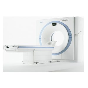 The Siemens Sensation 16 Slice CT is one of the most widely-used multi-slice, whole-body CT scanners on the market. With the ability to perform 3D whole body and cardiac exams with exceptional image quality, the Siemens Sensation 16 isan excellent choice for imaging centers without the budget to buy new. The Siemens Sensation 16-Slice CT matures RRealtime MPR CT Angio, 2D Post Processing, 3D Display, Syngo Shaded Display(SSD), Volume Measurements, and Volume Rendering Technique. Technology 42 Second Rotation Speed Dura Akron Tube – 5.3 MHU Navigator Console Wizard Work Station 18” Flat Screen Monitors Air Cooled System Syngo UserInterface
Siemens Sensation 16
-
 The ANATOM 64 CT scanner is the latest innovation for cardiac imaging based on Precision Platform system. The excellent design of Ahart technology which innovatively combined single spiral scan + gated imaging + mA modulation for easy heart imaging at extremely low radiation dose. We provide you ANATOM 64 Clarity/Precision of two models which are low/high configurations for preferences. It also offers you conventional clinical applications of low dose, better image quality and faster exams. Features: Modularized OptiWave HD detector features low-cost & easy maintenance, high spatial resolution and long lifetime Admir3D iterative technology delivers optimal dose efficiency and noise reduction without compromising image quality High configurations of main components ensure the best results and maximum patient throughput Uniquely and creatively uses 140kV and 80kV dual energy scan mode for brain imaging on 16-slice CT to offers you extraordinary image quality both in low and high density resolutions AdoseTM mA modulation ensures you low dose imaging without compromising image quality particularly useful in pediatric applications Equipped with dedicated Abast and Amast for bone and metal artifacts The brilliant Ahart technology enables you to experience so easy and low-dose cardiac imaging applications Technical Specifications: Model ANATOM 64 Precision ANATOM 64 Fit Rack type Low pressure slip ring Low pressure slip ring Scan aperture 70cm 70cm Rack Physical inclination ± 30 ° N.A Rack digital inclination ± 50 ° ± 50 ° cooling method Air-cooled Air-cooled Focus to the center distance 56 cm 53 cm Maximum power (non-equivalent) 80kW 42kW Votage (kV) 80kV / 100kV / 120kV / 140kV 70kV / 80kV / 100kV / 120kV / 140kV Heat capacity 8MHU 5.0MHU Heat dissipation rate 931 kHU / min 748kHU / min cooling method Oil cool Oil cool Large focus size 1.1mm × 1.2mm 1.2mm × 1.4mm Small focus size 0.6mm × 1.2mm 0.7mm × 0.8mm Tube current range 10-670mA 10-350mA Detector type Optiwave detectors Optiwave detectors Number of Z-axis 32 32 The width of the Z-axis 20mm 20mm The number of elements per row 912 848 Total number of detectors 29184 27136 Acquisition mode 64×0.625, 32×0.625, 16×0.625 64×0.625, 32×0.625, 16×0.625 Scanning range 1800mm 1800mm Horizontal positioning accuracy ± 0.25mm ± 0.25mm weight capacity 205kg 205kg Minimum height 43cm 43cm Anti – collision protection device Yes Yes Foot control switch Yes Yes IV rack Yes Yes CPU 3.5GHz 3.5GHz RAM 16 GB × 4 16 GB × 4 Hard drive capacity 1T × 2 1T × 2 Display size 24 inch LCD monitor 24 inch LCD monitor Display resolution 1920 × 1200 1920 × 1200 Computer operating system Windows 7 Windows 7 Image reconstruction speed 65 frames/ second 65 frames/ second Number of image store 1000000 1000000 Data external storage mode CD / DVD / USB CD / DVD / USB Minimum Scan Time of 360 degree 0.39sec 0.75sec Sub-millimeter acquisition layers 64 64 Double sub-millimeter acquisition layers 64 64 Thinnest acquisition thickness 0.625mm 0.625mm The thinnest reconstruction thickness 0.3125mm 0.625mm Conventional reconstruction thickness (mm) 0.3125 mm, 0.625 mm, 1.25 mm, 2.5 mm, 5.0 mm, 7.5 mm, 10 mm 0.625 mm, 1.25 mm, 2.5 mm, 5.0 mm, 7.5 mm, 10 mm The reconstruction matrix 512 x 512, 1024 x 1024 512 x 512, 1024 x 1024 Display matrix 1024 × 1024 1024 × 1024 Max FOV 52cm 50cm The maximum display field of view 70cm 50cm Maximum scan length 1800mm 1800mm Maximum continuous helix scan time 120s 120s Pitch range 0.5-1.5 0.5, 1.0, 1.5 High contrast resolution 21 Lp / cm @ 0% MTF 21 Lp / cm @ 0% MTF Low contrast resolution 2mm @ 0.3% 2mm @ 0.3% Image noise ≤ 0.25 ≤ 0.29 MPR Yes Yes CPR Yes Yes SSD Yes Yes VR Yes Yes MIP Yes Yes MinIP Yes Yes Virtual endoscopy Yes Yes CT angiography Yes Yes Tissue segmentation Yes Yes One-key bone removal Yes Yes Automatically patient table removal Yes Yes Contrast Agent Automatic Tracking Technology- bolus tracking Yes Yes Automatic linkage trigger technology Yes Yes Cine mode display Yes Yes Bone artifact suppression technique AbastTM AbastTM Metal artifact suppression technique AbastTM AbastTM Iterative reconstruction technique Admir3D global iteration Admir3D full-domain iteration Low – dose children ‘s scanning technology Yes Yes Low – dose lung scan Yes Yes Gray matter enhancement technology AccuHead AccuHead High resolution imaging of the lung AccuLung AccuLung Inner ear high resolution imaging AccuOtica AccuOtica Body high resolution imaging AccuBody AccuBody Bone high resolution imaging AccuBone AccuBone Head dual-energy imaging Ahead Ahead CT perfusion imaging Optional Optional Quantitative analysis of blood vessels Optional Optional Heart coronary artery imaging Aheart N.A ECG gated Yes N.A Low dose cardiac scan Yes N.A Green energy saving technology AccuSaving AccuSaving Dual-energy scan technology Optional Optional Click Here To Download Catalogue
Anke Anatom 64 Clarity Multi-Slice Spiral CT Scan
-
 This Machine gives a possibility to perform computed tomography without any problems and on high quality level. This device is used to conduct exams of internal organs and their functioning. With its help, a physician has a possibility to assess the condition of the human body as a whole. Features: It is easy to use; Convenience; Multi functionality; Obtained images are of high definition; High-definition 3D images of the area under study; The procedure is pain-free; The data is processed fast; The image can be stored in the computer memory; The diagnostics does not take a lot of time; Acceptable radiation dose. Technical Specifications: No. Technical Features s 1 Gantry 1.01 Gantry type Low voltage slip-ring 1.02 Gantry driven type Strap-driven 1.03 Patient opening 70cm 1.04 Gantry tilt mode Digital gantry tilt 1.05 Digital tilt capability ±50° 1.06 Detector type OptiWave rare-earth ceramic detector 1.07 Numbers of detector rows 16 1.08 Width of Z-axle detector 20mm 1.09 Detector columns of channels per row 848 1.10 Numbers of detector columns 13568 1.11 Data-transfer type RF, optical fiber communication 1.12 Distance of focus-ISO-center 53cm 1.13 Distance of focus-detector 94cm 1.14 3D laser orientation Provided 1.15 13″ integrated display panel Provided 1.16 Adose automatic exposure control (mA Modulation) Provided 1.17 Auto-voice manager Breath Graphical Display Hold Message (Record/Playback) Breath Message (Record/Playback) 1.18 AccuSaving energy conservation management Provided 2 HVPS and X-ray tube 2.01 Maximum continuous output of HVgenerator 42kW 2.02 Tube kV selections 70kV, 80kV, 100 kV, 120 kV, 140 kV 2.03 Tube mA range 10~350mA 2.04 Tube anode heat capacity 3.5MHU 2.05 Max. anode cooling rate 735kHU/min 2.06 Type of cooling Oil cooling + Air cooling 2.07 Tube focus Large: 1.2mm×1.4mm Small: 0.7mm×0.8mm 2.08 Collimator width selection 4-level election 2.09 Focus spot tracking technology Provided 3 Patient table 3.01 Maximum horizontal-movable range 1850mm 3.02 Table horizontal-scannablerange 1800mm 3.03 Table horizontal-position repeatability ±0.25mm 3.04 Minimum height above floor 430mm 3.05 Maximum vertical-movable range 500mm 3.06 Maximum speed of vertical movement 35mm 3.07 Maximum speed of horizontal movement 150mm/s 3.08 Maximum patient weight 205kg 3.09 Foot pedal of patient table control Provided 4 Computer 4.01 CPU 3.5GHz 4.02 Memory 32GB 4.03 Storage of hard-disk 1TB×2 4.04 Monitor 24’’ LCD Monitor 4.05 Resolution of monitor 1920×1200 4.06 Image-data external storage type CD/DVD/USB 4.07 Time of image reconstruction (512×512) 33.3ms/image 4.08 Speed of image reconstruction (512×12) 30fps 4.09 DICOM 3.0 interface Provided 4.10 Printer DICOM 3.0 interface Provided 4.11 Auto filming Provided 4.12 Worklist function Provided 5 Scan parameters 5.01 Shortest 360 degree rotation time 0.75s 5.02 Allowed rotation times 0.75s, 1.0s, 1.5s, 2.0s, 3.0s, 4.0s 5.03 Maximum slice numbers per rotation 32 5.04 Minimum slice thickness of scan 1.25mm 5.05 Minimum slice thickness of reconstruction 0.625mm 5.06 Maximum slice thickness of scan 20mm 5.07 Nominal reconstruction slice thickness 0.625mm, 1.25mm, 2.5mm, 5.0mm, 7.5mm, 10mm, 20mm 5.08 Speed of image reconstruction (512×512) 30 frames/s 5.09 Scan FOV 50cm 5.10 Image reconstruction matrix 512×512, 1024×1024 (Optional) 5.11 Image reconstruction matrix 512×512, 1024×1024 (Optional) 5.12 Image display matrix 512×512, 1024×1024 (Optional) 5.13 Maximum continuous scan duration 120s 5.14 Maximum continuous scan length 180cm 5.15 Direction of TOPO Front-back, Left-right 5.16 Max. length of TOPO 180cm 5.17 Range of pitch 0.5~1.5 5.18 Scan mode Scout scan Axial scan Helical scan Cine scan 6 Image Quality 6.01 High contrast resolution 21lp/cm@0%MTF 6.02 Low contrast resolution 2.0mm@0.30% 6.03 Isotropic imaging resolution 0.24mm 6.04 Range of CT numbers -32767~32768 6.05 Image noise ≤0.29@28mGy 7 Advanced application 7.01 Multi-Planar Reconstruction (MPR) Provided 7.02 Curve Multi-Planar Reconstruction (CPR) Provided 7.03 Surface Shaded Display (SSD) Provided 7.04 Volume Rendering (VR) Provided 7.05 Maximum Intensity Projection (MIP) Provided 7.06 Minimum Intensity Projection (MinIP) Provided 7.07 Virtual Endoscopy (VE) Provided 7.08 CT angiography (CTA) Provided 7.09 Tissue segmentation Provided 7.10 One click bone remove Provided 7.11 One click patient table remove Provided 7.12 Bolus-tracking Technology Provided 7.13 Spiral auto start Provided 7.14 Cine display Provided 7.15 AbastTM bone artifact suppression technology Provided 7.16 AmastTM metal artifact suppression technology Provided 7.17 Admir3D all-domain iterative reconstruction Provided 7.18 Low-dose pediatric scan technology Provided 7.19 Low-dose lung scan technology Provided 7.20 AccuHead grey-white matter enhanced technology Provided 7.21 AccuOrgan lung high resolution scan technology Provided 7.22 AccuOrgan inner-ear high resolution scan technology Provided 7.23 AccuOrgan body high resolution scan technology Provided 7.24 AccuOrgan bone high resolution scan technology Provided 7.25 AccuMatter dual-energy with Admir3D for new application Provided Click Here To Download Catalogue
Anke Anatom 32 Fit Multi-Slice Spiral CT Scan
-
 ANATOM16 HD, is a tool of precision medicine in diagnosis imaging. Via the breakthrough designs in precise hardware, software and imaging technologies, ANATOM 16 HD can provide precise diagnosis information and early detection for small lesions. Features: Seamlessly upgrade to meet your future needs: Anke takes full consideration of the increasing clinical requirements of your business in today’s rapidly changing medical environment. Precise hardware, Precise technology, Precise imaging: OptiWave detector, High precision gantry control, Dual-mode gantry tilt, Admir3D iterative technology, Dual-energy head imaging, 1024 x1024 matrix imaging technology, High-definition imaging of targeted organs, Low dose platform, 3D enhanced VR Precision Technology Platform: ANATOM precision technology platform is equipped with advanced imaging technologies, and adopts OptiWave detector, Ahead dual-energy imaging, Admir3D iterative reconstruction technology and AccuTilt dual-mode tilt gantry technology to provide powerful support for accurate diagnosis Admir3D iterative reconstruction technology: Admir applies mathematical and physics models to accurately construct and describe the signal’s quantum characteristics. Iterative operations are performed in the three domains of raw data, projection and image, to greatly reduce the image noise and achieve optimal image quality with low dose Ahead-Head dual-energy head imaging technology: Ahead creatively uses 140kV and 80kV dual energy switching scan mode for brain imaging. By careful analyzing the high and low energy characteristics, images can show more valuable information about the brain tissues AccuTilt dual-mode gantry tilt technology: The system provides digital and mechanical tilt to accommodate different user habits and clinical needs. Real-time collision preventing system is available for the patients’ safety AccuOrgan-Targeted organ imaging: To achieve high precision imaging of each part of human body at low dose and low energy consumption AccuDose-Comprehensive low dose imaging: Pediatric Scan Protocol, Individual Dose Monitoring, AccuShape Filter, Efficient Detector, Adose Dose Modulation, Ahead – Head Dual-energy Imaging, Iterative Reconstruction, Amast, Contrast Agent Tracking Technology AccuScan-Enjoy ease: Convenient and efficient operation process greatly improve work efficiency to achieve high volume of patients Clinical Applications: Fast, precise and low-dose imaging technologies provide a full range of clinical solutions to meet the current and future clinical diagnostic needs Service Innovation creating maximum value for customers: Service Support within 24 Hours, Local Service Partners, On-line Service Support, After-sales Maintenance Stations AccuSaving Green & Energy-saving: AccuSaving is an innovative energy saving technology. The system will enter the “dormant”, which is a low carbon mode, after a certain idle time or per user’s request. To bring the system back to working status is as easy as pushing a button. The system will also remind the user to perform necessary warm-up and calibration procedures, which are fully automated processes. AccuSaving technology can reduce operation and standby power consumption and save the electricity cost by 30% by adopting different operation modes in working and off hours Technical Specifications: No. Technical feature 1 Gantry 1.01 Gantry type Low voltage slip-ring with AccuSlip-ring technology 1.02 Gantry driven type Strap-driven 1.03 Patient opening 70cm 1.04 Gantry tilt mode Dual-mode gantry tilt 1.05 Mechanical tilt capability ±30° 1.06 Digital tilt capability ±50° 1.07 Gantry remote-Control Provided 1.08 Detector type OptiWave rare-earth ceramic detector 1.09 Numbers of detector rows 32 1.10 Width of Z-axle detector 20mm 1.11 Detector columns of channels per row 912 1.12 Numbers of detector columns 29184 1.13 Data-transfer type RF,optical fiber communication 1.14 3D laser orientation Provided 1.15 External X-ray enable Interface for Foot-Pedal Provided 1.16 Automatic exposure control(mA Modulation) Provided 1.17 Auto-voice manager Breath Graphical Display Hold Message (Record/Playback) Breath Message(Record/Playback) 1.18 ANKE energy conservation management Provided 1.19 Acquisition mode 16 × 0.625mm, 16 × 1.25mm 2 Scan parameter 2.01 Shortest 360 degree rotation time 0.5s 2.02 Allowed rotation times 0.5s,0.8s,1.0s,1.5s,2.0s 2.03 Slice numbers per rotation 16 2.04 Minimum slice thickness of scan 0.625mm 2.05 Minimum slice thickness of reconstruction 0.625mm 2.06 Maximum slice thickness of scan 10mm 2.07 Nominal reconstruction slice thickness 0.625mm,1.25mm,2.5mm,5.0mm, 7.5mm,10mm 2.08 Speed of image reconstruction(512×512) 65 frames/s 2.09 Scan FOV 52cm 2.10 Image reconstruction matrix 512×512,1024×1024 2.11 Image display matrix 512×512,1024×1024 2.12 Maximum continuous scan duration 120s 2.13 Maximum continuous scan length 180cm 2.14 Direction of TOPO Front-back,Left-right 2.15 Max. length of TOPO 180cm 2.16 Range of pitch 0.5~1.5 2.17 Scan mode Scout scan Axial scan Helical scan Cine scan 3 HVPS and Tube 3.01 Maximum continuous output of HV generator 50kW 3.02 Tube kV selections 80kV,100 kV,120 kV,140 kV 3.03 Tube mA range 10~420mA 3.04 Tube anode heat capacity 5.0MHU 3.05 Heat dissipation rate 815kHU/min 3.06 Type of cooling Oil cooling + Air cooling 3.07 Tube focus Large:1.0 mm×1.0mm Small:0.5mm×1.0mm 3.08 Dynamic flying focal spot technology Provided 4 Patient table 4.01 Maximum horizontal-movable range 1850mm 4.02 Table horizontal-scannable range 1800mm 4.03 Table horizontal-position repeatability ±0.25mm 4.04 Maximum vertical-movable range 500mm 4.05 Maximum speed of vertical movement 20mm/s 4.06 Maximum speed of horizontal movement 150mm/s 4.07 Maximum patient weight 205kg 4.08 Foot pedal of patient table control Provided 5 Image Quality 5.01 High contrast resolution 21lp/cm@0%MTF 5.02 Low contrast resolution 2.0mm@0.30% 5.03 Isotropic imaging resolution 0.625mm 5.04 Range of CT numbers -32767~32768 5.05 Image noise ≤0.25@28mGy 6 Computer subsystem 6.01 CPU 3.5GHz 6.02 Memory 16GB×4 6.03 Storage of hard-disk 1T×2 6.04 Monitor 24’’ LCD Monitor 6.05 Resolution of monitor 1920×1200 6.06 Image-data external storage type CD/DVD/USB 6.07 Time of image reconstruction(512×512) 15.4ms/frame 6.08 DICOM 3.0 interface Provided 6.09 Printer DICOM 3.0 interface Provided 6.10 Auto filming Provided 6.11 Worklist function Provided 7 Advanced application 7.01 Multi-Planar Reconstruction(MPR) Provided 7.02 Curve Multi-Planar Reconstruction(CPR) Provided 7.03 Surface Shaded Display(SSD) Provided 7.04 Volume Rendering(VR) Provided 7.05 Maximum Intensity Projection(MIP) Provided 7.06 Minimum Intensity Projection(MinIP) Provided 7.07 Virtual Endoscopy(VE) Provided 7.08 CT angiography(CTA) Provided 7.09 Tissue segmentation Provided 7.10 One click bone remove Provided 7.11 One click patient table remove Provided 7.12 Bolus-tracking Technology Provided 7.13 Spiral auto start Provided 7.14 Cine display Provided 7.15 AbastTM bone artifact suppression technology Provided 7.16 AmastTM metal artifact suppression technology Provided 7.17 Admir3D fulll-domain iterative reconstruction Provided 7.18 Low-dose pediatric scan technology Provided 7.19 Low-dose lung scan technology Provided 7.20 AccuHead grey-white matter enhanced technology Provided 7.21 AccuLung high resolution scan technology Provided 7.22 AccuOtica inner-ear high resolution scan technology Provided 7.23 AccuBody high resolution scan technology Provided 7.24 AccuBone high resolution scan technology Provided Click Here To Download Catalogue
Anke Anatom 16 Slice CT Scan
-
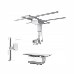 DrGem Ceiling Analogue X-ray Machine is a diagnostic radiography system X-ray Machine that provides reliable high quality radiographic images with a reduced dose. The reliable high-frequency x-ray generators that are known worldwide for their excellent performance, lifetime and stability. Patient tables and wall stands are also offered. Features of DrGem Ceiling Analogue X-ray Machine TS-CSA-A (Vertical movement, 1.6m stroke, rail length 3x4meter) including HV cable 15m WBS-TA: Vertical movement V Stroke:1,450mm in Uprigh Bucky Position, 1,526mm in Horizontal Bucky position. PBT-4 is a 4 way Floating Tabletop with. A large tabletop with extended travel enables all radiography studies with minimal patient movement. Fully fat tabletop without a frame on the edge makes cleanliness and odors free Technical Specifications of DrGem Ceiling Analogue X-ray Machine Power Rating – 32KW Generator – GXR-32S Rotor – Dual Speed Starter(DSS) Input Power – 400/480VAC, Three phase Line Frequency – 50/60Hz X-ray tube – DXT-12M, (0.6/1.2mm, 300kHU) Tube Voltage – 40 to 150kV, 1kV Step Tube Current – 10 to 640mA Output – 640mA@81kV, 500mA@104kV, 400mA@130kV, 320mA@150kV Time Range – 1ms to 10s mAs Range – 0.1 to 800mAs Reproducibility – Coecient of Variation : kV < 0.005, Time < 0.005,mAs < 0.01 Accuracy – kV < ±(1%+1kV), mA < ±(3%+1mA), Time <±(1%+0.5ms), mAs < ±(3%+0.1mAs) Linearity – Coecient of Linearity < 0.01 : CL = (X1-X2)/(X1+X2), where X is mR/mAs Mechanical Parts: -TS-CSA-A (Vertical movement, 1.6m, stroke rail length 3x4meter) including HV cable 15m – PBT-4: 4 way Floating Tabletop with Elevating Feature (66cm). – WBS-TA: a. Vertical movement V Stroke:1,450mm in Upright Bucky Position, 1,526mm in Horizontal Bucky position. – HVC-15: 15M HV cable – Auto Collimator Click Here To Download Catalogue
DrGem Ceiling Analogue X-ray Machine
Zenrox Healthcare Solutions - Advanced Medical Equipment in Lagos
Zenrox offers cutting-edge medical equipment and comprehensive support services in Lagos. Explore our products designed for superior healthcare delivery. Contact us today for innovative medical solutions.