-
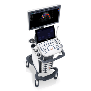
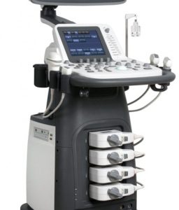
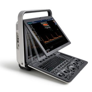
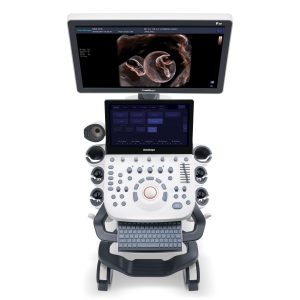 DETAILS Upgraded Images with More Clarity SonoScape never stops making progress in improving the image quality of its ultrasound products to enhance the confidence of diagnosis for doctors. With extraordinary images provided by P20, the anatomy structures are clearer than ever. C-Xlasto Imaging With C-xlasto Imaging, P20 enables comprehensive quantitative elastic analysis. Meanwhile, C-xlasto on P20 is supported by linear, convex and transvaginal probes, to ensure good reproducibility and highly consistent quantitative elastic results. S-Live S-Live allows for detailed visualization of subtle anatomical features, thereby enabling intuitive diagnosis with real-time 3D images and enriching patient communication. Pelvic Floor 4D Transperineal 4D pelvic floor ultrasound can provide useful clinical values in assessing the vaginal delivery impact on the female anterior compartment, judging whether the pelvic organs are prolapsed or not and the extent, determining if the pelvic muscles were torn accurately. Anatomic M Mode Anatomic M Mode helps you observe the myocardial motion at different phases by freely placing sample lines. It accurately measures the myocardial thickness and the heart size of even difficult patients and supports the myocardial function and LV wall-motion assessment. Tissue Doppler Imaging P20 is endowed with Tissue Doppler Imaging which provides velocities and other clinical information on myocardial functions, facilitating clinical doctors with the ability to analyze and compare the motions of different parts of the patient’s heart. Click Here To Download Catalogue
Sonoscape P20 Ultrasound Machine
-
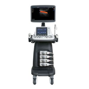
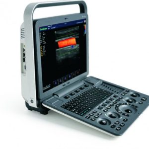
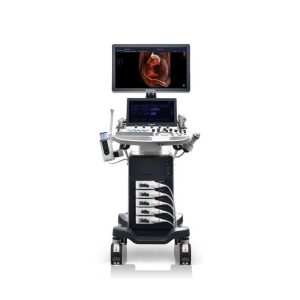 DETAILS Powerful Compact Precision Taking into consideration the evolving expectations and needs for ultrasound, the P50 is a slim and unobtrusive trolley system that is comfortable in tight, congested spaces with little room to work in. Providing everything you need for a comfortable examination in a small space for both you and your patient. Single Crystal Transducer Wideband single crystal probes greatly improve the signal ratio, acquire stunning images and provide superior sensitivity and resolution for both the near and far-fields. μ-Scan+ The new generation μ-Scan imaging technologies give you better image quality by reducing noise, improving signal strength and improving visualization. Dynamic Color Dynamic colour improves upon already existing colour Doppler technologies for clear capture of colour flow and detail visualization of even tiny veins with lower velocities. Solution for Radiology P50, is a leading-edge ultrasound system that can meet the demands of any clinical setting. You can experience a superior performance in multi-dimensional imaging for a full range of clinical applications – abdominal, breast and cardiovascular. C-xlasto Imaging By understanding that tissue stiffness varies depending on the type of tissue, we can use C-xlasto Imaging to easily find abnormalities and tumours within soft tissue. The differences in tissue responses are detected and visualized in real-time by the elastography algorithms through different representations, which can be particularly helpful in analyzing breast, thyroid and musculoskeletal structures. Predominately used only in linear probes, SonoScape’s new transvaginal and bi-plane probe for gynaecology and urology are breaking the mould and expanding elastography applications. Real-time Color Panoramic With the combination of colour flow and real-time panoramic, visualizing the blood flow of an entire vein or artery is now an easy task. Accomplished in real-time for the convenience of the sonographers, any mistakes can also be easily backtracked and corrected without interrupting the scan. Contrast Imaging Contrast Imaging on P50 makes full use of the infra harmonic signal and second harmonic signal to improve the image resolution and deep penetration. What’s more, the Dynamic Acoustic Control technology effectively controls the acoustic pressure for the contrast agent, decreasing the required agent dose and assures uniform image quality, guaranteeing longer contrast agent duration and better lesion perfusion of delayed phase observation. Solution for OB/GYN P50 has superior image quality, automated measurement tools, and a variety of volume technologies to provide ideal solutions for clinical examinations such as pregnancy examinations, and gynecologic disease diagnosis. With a new 4D transvaginal probe, P50 helps you to see and detect fetal abnormalities and significantly improves your diagnostic confidence during your examinations. S-Live Silhouette A unique transparent 3D anatomical image of the fetus for improved initial anatomical review. By using this new application, the system can create completely different fetal images from conventional ultrasound images, which can depict the fetal’s intracorporeal anatomical structure. Pelvic Floor 4D Working in conjunction with SonoScape’s latest transvaginal probes, trans-perineal 4D pelvic floor ultrasound provides a useful clinical assessment of the impact of vaginal delivery on the female anterior compartment. Allowing doctors to judge whether the pelvic organs prolapsed or not, the extent of prolapse, and determining whether the pelvic muscles tore correctly. S-Guide S-Guide gives the user an extensive list of example obstetric ultrasound images as reference guides and a convenient checklist system to keep track of their progress during their obstetrics examination. Auto Face Automatically removes masking layers in front of the fetus’s face for a clearer vision of the fetus’s face. AVC Follicle AVC Follicle automatically identifies how many follicles are present and calculates their individual volumes. Solution for Cardiology P50 provides clear 2D clinical images and Doppler sensitivity to assess critical cardiac performance. Compatible with SonoScape’s single crystal probes, the P50 can provide images with better resolution and penetration in Cardiac diagnosis. Tissue Doppler Imaging Tissue Doppler Imaging allows clinical doctors to quantitatively evaluate local myocardial movements and functions, facilitating them with the ability to analyze and compare the motions of the different parts of the patient’s heart. Stress Echo Stress echocardiography is the combination of 2D echocardiography with physical, pharmacological or electrical stress of the patient. It also then provides users with report management tools such as configurable template editor, multiple loops to select one for storage, wall motion scoring, stress echo report, etc Auto IMT Auto IMT is used when determining the level of vascular sclerosis present in the patient by automatically tracing and calculating the thickness of the carotid vessels. What distinguishes the P50 is that it provides an instant and accurate Mean and Max index at the touch of a single button. Auto EF Automated 2D Cardiac Quantification is a fully intelligent trace function for endocardium with 19 easily-adjustable points providing rapid access to proven 2D EF and volumes. Click Here To Download Catalogue
Sonoscape P50 Ultrasound Machine
-
 15” high definition LED monitor with articulating arm Four transducer sockets A comprehensive selection of probes: Linear, Convex, Micro-convex, Endocavity, Phased Array, Bi-plane, Volumetric Premium application technology: μ-scan speckle reduction, Pulse Inversion Harmonic Imaging, Realtime Panoramic Imaging, Trapezoid Imaging, AutoIMT, Triplex Full patient database and image management solutions DICOM 3.0, AVI/JPG, USB2.0, HDD, DVD, PDF report Built-in Battery
Sonoscape S12 Ultrasounds
-
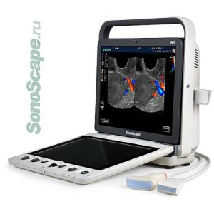
 15-inch high resolution LCD monitor with wide-view angle Standard PC keyboard, easy input Two probe sockets with probe holder, better protection for probes Rechargeable lithium battery, 1 hour scanning without power supply Abundant peripherals: DICOM3.0, VGA, video out, USB, S-Video, ECG module, Foot switch etc. B Mode, Dual B, 4B M Mode, Color M, Steer M,SonoScape S2 Front Color Mode, DPI Mode PW Mode, CW Mode (optional) Advanced Imaging Technology THI μ-Scan Speckle Reduction (optional) Compound Imaging (optional) Panoramic Imaging (optional) Trapezoid Imaging (optional) 4D imaging (optional)
Sonoscape S2 Ultrasound
-
 18.5″ high resolution widescreen LED 8″ touch screen Premium application technology: I4D, μ-scan, com pound imaging, Pulse Inversion Harmonic Imaging, Color M-mode, Steer M-mode, PDI, TDI, Real-time Panoramic, Trapezoid, Auto-IMT, Stress Echo Full patient database and image management Solutions: DICOM 3.0, AVI / JPG, USB2.0, HDD, DVD, PDF report Multi-language Input Keyboard Mode B-mode, M-mode, THI, CDI, DPI, TDI, PW, CW, HPRF, 3D / 4D, Color M-mode, Steer Mmode, Panoramic imaging Scan format Linear, Convex, Micro-convex, Endocavity, Phased Array, Intraoperative, TEE, Biplane, Pencil, Volumetric Transducer inputs 4 + 1
Sonoscape S22 Ultrasound
-
 DETAILS As SonoScape steps forward to add value and efficiency to ultrasound, the latest S22 was designed in a user-friendly platform to address current and future demanding needs. It represents an excellent mix in performance and price. S22, is a shared service ultrasound system with a slim and elegant package that has combined mobility with utility to fit in specific clinical situations including emergency department, ICU, operating room and so on. Furthermore, its ergonomic design, easy operating and flexible data management will give you a memorable experience. SPECIFICATION • Large high-resolution widescreen LED • Sensitive touch screen • Four transducer sockets plus one socket for pencil probe • A comprehensive selection of probes: linear, Convex, Micro-convex, Volumetric, Endocavity, Bi-plane, Phased Array, TEE, Intraoperative, Pencil • Premium application technology: 4D, μ-scan speckle reduction, compound imaging, Pulse Inversion Harmonic Imaging, Color M-Mode, Steer M-Mode, PDI, TDI, Real-time Panoramic Imaging, Trapezoid Imaging, Auto-IMT… • Full patient database and image management solutions: DICOM 3.0, AVI/JPG, USB 2.0, HDD, DVD, PDF report • Multi-Language Input Keyboard • Built-in battery Click Here To Download Catalogue
Sonoscape S22 Ultrasound Machine
-
 S40 Technology and Elegance 19 inch definition LCD monitor with wide viewing angle 10 inch touch screen with 15°adjustable angle Height and position adjustable control plain with muted function Five transducer sockets and additional one for CW probe Additional Endocavity probe holder and gel warmer Full range of transducers: Linear, Convex, Micro-convex, Endocavity, Phased array, Intraoperative, TEE, Pencil probe, Volumetric, Endocavity 4D and Laparoscope probe Advanced application technology: TDI, Stress Echo and Elastography Full patient database solution: DICOM 3.0, AVI/JPG, Dual USB, HDD, DVD, PDF report
Sonoscape S40 Ultrasound
-
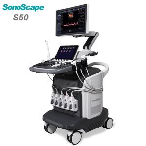 The design of s50 took operational use into consideration, creating a comfortable diagnosing enviroment. Ergonomic design, excellent man-machine interaction and rapid response, makes s50 an intelligent scanning assistant for you, bringing improved efficiency and helping to prevent fatigue from multiple examinations. 21.5 inches wide Led Monitor Super Responsive Touch Screen Flexible Adjusting Control Panel Built in Gel Warmer Wireless Wi-Fi Connection
Sonoscape S50 Ultrasound
-
Hot
 Sonoscape S8 Exp Portable Ultrasound scanner DETAILS Agile and Versatile With ultra-modern innovative design and the clinically-proven technologies, S8 Exp Portable Ultrasound scanner is well equipped as a low-physical-effort and enhanced-image-quality ultrasound scanner, which not only provides optimized solutions for versatile applications but does help to improve the user experience for both routine and non-traditional challenges. Working with S8 Exp, it will trigger your unlimited reverie and endow you with endless charm. Carrying forward the classical design of SonoScape’s portable ultrasound products, S8 Exp successfully combines the best ergonomics, attractive design and ease of use. This charismatic identity is also enhanced by a sophisticated color palette—with sedate grey as its interior paint color and pearl white as exterior cover, S8 Exp reveals a style of aristocrat and strong character among SonoScape’s ultrasound systems. Workflow The S8 Exp is a portable ultrasound scanner that adapts to your workflow, whether you are in the consulting room, at the bedside, or at a remote location. With easy-to-use new platform designed for sonographers’ needs and full connection interfaces for easy connectivity and data sharing, S8 Exp leads to improved user comfort and clinical outcome as well as patient throughput and working efficiency. Powerful Platform Embedded with SonoScape‘s core imaging technologies such as μ-scan, PHI and Spatial Compound, S8 Exp boasts exceptional 2D image, sensitive spectral, Color and Power Doppler, displaying well-defined anatomy and pathology and facilitating a highly optimized diagnostic user environment for conclusive diagnoses. Besides, S8 Exp offers a comprehensive selection of electronic probes to maximally extend its capabilities to meet a wide range of applications including the abdomen, pediatric, OB/GYN, cardiovascular, musculoskeletal, etc. The advanced probe technologies also effectively enhance the image quality and confidence in reaching clinical diagnoses even in difficult patients. Click Here To Download Catalogue
Sonoscape S8 Exp Portable Ultrasound
-
 LCD 15” Monitor angle adjustment 2 active probe connectors High density probes Scanning modes: В, М, В/М, В/В, 4В High precision tissue harmonics ZOOM at real time and frozen state Ultrasound tomography mode TRAPEZOIDAL mode at linear probes MicroScan speckle reduction imaging Color Doppler mode Power Doppler mode Directional power Doppler mode Pulse wave Doppler mode Continuous Doppler mode Color tissue Doppler mode Pulse wave tissue Doppler mode Duplex and Triplex modes Steer M-mode Color M-mode Free Hand 3D mode 4D mode PANORAMIC mode ELASTO with quantitative evaluation STRESS ECHO mode
Sonoscape S8EXP Ultrasound
-
 Modern technologies, innovative design and perfect imaging provide unlimited opportunities to the S9 system users LCD 15” Monitor angle adjustment 2 active probe connectors Touch control panel High density probes Scanning modes: В, М, В/М, В/В, 4В High precision tissue harmonics ZOOM at real time and frozen state Ultrasound tomography mode TRAPEZOIDAL mode at linear probes MicroScan speckle reduction imaging Color Doppler mode Power Doppler mode Directional power Doppler mode Pulse wave Doppler mode Continuous Doppler mode Color tissue Doppler mode Pulse wave tissue Doppler mode Duplex and Triplex modes Steer M-mode Steer M-mode Color M-mode Free Hand 3D mode 4D mode Panoramic Mode Elasto With Quantitative Evaluation Stress Echo Mode
Sonoscape S9 Ultrasound
-
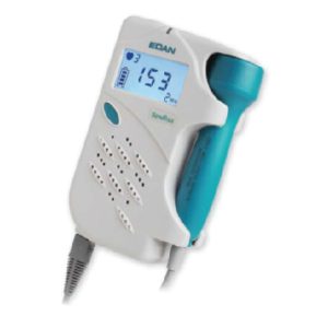 SonoTrax Series Ultrasonic Pocket Doppler provides caregivers with the most cost effective solution to meet the routine obstetric and vascular requirements. Easy to use Compact design with easy one-hand operation Bright LED screen with real-time FHR display 2 MHZ probe with deep penetration for later pregnancy and larger patients 3 MHZ probe detects fetal heart beat as early as 9 weeks 4/5/8MHz probe for blood flow detection Real-time monitoring for daily use Record & play with audio playback and numeric display Built-in Li-ion rechargeable battery ensures long working time Unique charger stand design
SonoTrax Series Ultrasonic Pocket Doppler
-
Feature
 Innovative User-friendly Design
- 360°rotation of the X-ray tube column for any special radiographic positions.
- Pluggable detector and grid design.
Functional Applications- Automatic Exposure Control(AEC) function(Optional).
- Standard Dose Area Product(DAP) function.
- Pneumoconiosis Anatomic Procedural Radiography (APR) provides chest X-ray screening for pneumoconiosis.

 Stable Input Power Guarantee
- SONTU Power Solutions break limited input power with a single-phase option available (Optional). Excellent performance under various electrical and installation circumstances.
Multiple Configurations- Providing 50kW and 63kW high-frequency generator options.
- 14*17/17*17, wired/wireless flat panel detectors available.

Clinical Image
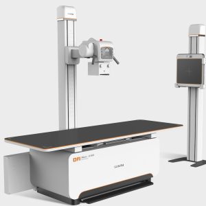
Sontu Statics X Ray sontu-100rad RAD(S) Series Floor-mounted DR
-
Hot
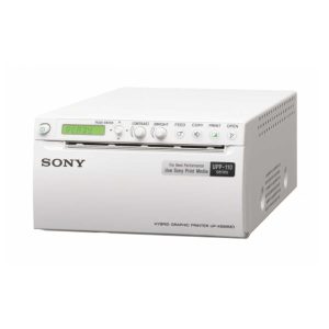 The UP-X898MD is a compact A6 medical grade black and white digital printer that can accept analogue video as well as PC-friendly digital USB signal inputs. It’s designed to be integrated into a wide range of compatible medical imaging environments such as digital ultrasound systems, mobile C-arms or cardiac catheterisation laboratory. The high quality thermal print engine can produce hard copy prints of still images captured by these systems for use in patient records and referrals. For even greater convenience, printed still images can also be stored to a connected USB flash drive. Features: High print quality A6 prints in less than two seconds Hybrid operation with analogue and digital inputs Compact, space-saving design Hard copy reference of print settings Wide PC compatibility Wireless Printing Capability Dual cutter configuration Store pictures on a USB flash drive Enhanced operability Uses widely available print media Quick guide print Compliance with Medical Safety Standards Technical Specifications: Printing Method: Direct thermal printing Resolution: 325 dp Gradations: 8 bits (256 levels) processing Picture Elements: Digital: 4096 x 1280 dots Video NTSC: 720 x 504 dots Video PAL: 720 x 604 dots Picture Area: Digital: 320 x 100 mm/12 5/8 x 3 7/8 inch (Max) STD Video NTSC: 94 x 73 mm, Video PAL: 94 x 71 mm (WIDE1) when “SIDE:OFF” is selected SIDE Video-NTSC: 124 x 96 mm, Video-PAL: 127 x 96 mm (WIDE1) when “SIDE:ON” is selected Paper Size: Paper width of 110 mm (4 3/8 inches) Printing Time: High-speed mode: Approx. 1.9 seconds/image (at standard setting) Normal speed mode: Approx. 3.3 seconds/image (at standard setting) Picture Memory: Digital: 4,096 x 1,280 x 8 (bit) Video: 10 frame memories (850 k x 8 bits per frame) Interface: USB terminal (type A) for USB flash drive (X1) Hi-Speed USB (USB 2.0) (x1) VIDEO INPUT: BNC type (x1) NTSC or PAL composite video signals 1.0 Vp-p, 75 ohms (NTSC/PAL automatically discriminated) VIDEO OUTPUT: BNC type (x1) Loop-through REMOTE: Stereo mini jack (x1) Printer Driver Software: Microsoft Windows 7 32/64 bit, 8 32/64 bit, 10 32/64 bit Power Requirements: AC 100 V to 240 V, 50/60 Hz Input Current: 1.3 A to 0.6 A Operating Temperature: 5°C to 40°C (41°F to 104°F) Operating Humidity: 20% to 80% (no condensation allowed) Storage and Transport Temperature: -20°C to +60°C (-4°F to +140°F) Storage and Transport Humidity: 20% to 80% (no condensation allowed) Dimensions (W x H x D): 154 x 88 x 240 mm (6 1/6 x 3 1/2 x 9 1/2 inches) Mass: 2.5 kg (5 lb 8 oz) Supplied Accessories: Thermal head cleaning sheet (1), CD-ROM (1), Before Using this Printer (1), Service Contact List (1), USB Flash Drive Ex. Cable, Print media (UPP-110HG)
Sony Thermal Printer
-
Hot
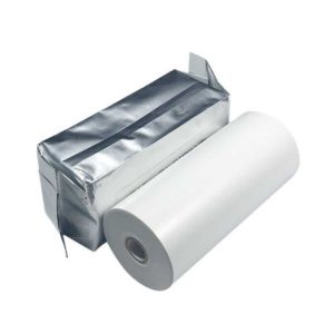 This A6 width B&W High Quality print paper (type I) is for use in UP-X898MD / D898MD / D898DC / 897MD / D897 / 895 / D895 / 890 / D890 / 860 / D860 printers. Mainly used in Ultrasound, Dental and Microscopy applications.
Sony Thermal Printer Paper
Zenrox Healthcare Solutions - Advanced Medical Equipment in Lagos
Zenrox offers cutting-edge medical equipment and comprehensive support services in Lagos. Explore our products designed for superior healthcare delivery. Contact us today for innovative medical solutions.