-
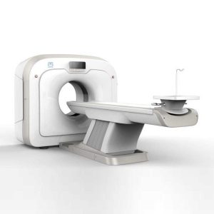
 ANATOM16 HD, is a tool of precision medicine in diagnosis imaging. Via the breakthrough designs in precise hardware, software and imaging technologies, ANATOM 16 HD can provide precise diagnosis information and early detection for small lesions. Features: Seamlessly upgrade to meet your future needs: Anke takes full consideration of the increasing clinical requirements of your business in today’s rapidly changing medical environment. Precise hardware, Precise technology, Precise imaging: OptiWave detector, High precision gantry control, Dual-mode gantry tilt, Admir3D iterative technology, Dual-energy head imaging, 1024 x1024 matrix imaging technology, High-definition imaging of targeted organs, Low dose platform, 3D enhanced VR Precision Technology Platform: ANATOM precision technology platform is equipped with advanced imaging technologies, and adopts OptiWave detector, Ahead dual-energy imaging, Admir3D iterative reconstruction technology and AccuTilt dual-mode tilt gantry technology to provide powerful support for accurate diagnosis Admir3D iterative reconstruction technology: Admir applies mathematical and physics models to accurately construct and describe the signal’s quantum characteristics. Iterative operations are performed in the three domains of raw data, projection and image, to greatly reduce the image noise and achieve optimal image quality with low dose Ahead-Head dual-energy head imaging technology: Ahead creatively uses 140kV and 80kV dual energy switching scan mode for brain imaging. By careful analyzing the high and low energy characteristics, images can show more valuable information about the brain tissues AccuTilt dual-mode gantry tilt technology: The system provides digital and mechanical tilt to accommodate different user habits and clinical needs. Real-time collision preventing system is available for the patients’ safety AccuOrgan-Targeted organ imaging: To achieve high precision imaging of each part of human body at low dose and low energy consumption AccuDose-Comprehensive low dose imaging: Pediatric Scan Protocol, Individual Dose Monitoring, AccuShape Filter, Efficient Detector, Adose Dose Modulation, Ahead – Head Dual-energy Imaging, Iterative Reconstruction, Amast, Contrast Agent Tracking Technology AccuScan-Enjoy ease: Convenient and efficient operation process greatly improve work efficiency to achieve high volume of patients Clinical Applications: Fast, precise and low-dose imaging technologies provide a full range of clinical solutions to meet the current and future clinical diagnostic needs Service Innovation creating maximum value for customers: Service Support within 24 Hours, Local Service Partners, On-line Service Support, After-sales Maintenance Stations AccuSaving Green & Energy-saving: AccuSaving is an innovative energy saving technology. The system will enter the “dormant”, which is a low carbon mode, after a certain idle time or per user’s request. To bring the system back to working status is as easy as pushing a button. The system will also remind the user to perform necessary warm-up and calibration procedures, which are fully automated processes. AccuSaving technology can reduce operation and standby power consumption and save the electricity cost by 30% by adopting different operation modes in working and off hours Technical Specifications: No. Technical feature 1 Gantry 1.01 Gantry type Low voltage slip-ring with AccuSlip-ring technology 1.02 Gantry driven type Strap-driven 1.03 Patient opening 70cm 1.04 Gantry tilt mode Dual-mode gantry tilt 1.05 Mechanical tilt capability ±30° 1.06 Digital tilt capability ±50° 1.07 Gantry remote-Control Provided 1.08 Detector type OptiWave rare-earth ceramic detector 1.09 Numbers of detector rows 32 1.10 Width of Z-axle detector 20mm 1.11 Detector columns of channels per row 912 1.12 Numbers of detector columns 29184 1.13 Data-transfer type RF,optical fiber communication 1.14 3D laser orientation Provided 1.15 External X-ray enable Interface for Foot-Pedal Provided 1.16 Automatic exposure control(mA Modulation) Provided 1.17 Auto-voice manager Breath Graphical Display Hold Message (Record/Playback) Breath Message(Record/Playback) 1.18 ANKE energy conservation management Provided 1.19 Acquisition mode 16 × 0.625mm, 16 × 1.25mm 2 Scan parameter 2.01 Shortest 360 degree rotation time 0.5s 2.02 Allowed rotation times 0.5s,0.8s,1.0s,1.5s,2.0s 2.03 Slice numbers per rotation 16 2.04 Minimum slice thickness of scan 0.625mm 2.05 Minimum slice thickness of reconstruction 0.625mm 2.06 Maximum slice thickness of scan 10mm 2.07 Nominal reconstruction slice thickness 0.625mm,1.25mm,2.5mm,5.0mm, 7.5mm,10mm 2.08 Speed of image reconstruction(512×512) 65 frames/s 2.09 Scan FOV 52cm 2.10 Image reconstruction matrix 512×512,1024×1024 2.11 Image display matrix 512×512,1024×1024 2.12 Maximum continuous scan duration 120s 2.13 Maximum continuous scan length 180cm 2.14 Direction of TOPO Front-back,Left-right 2.15 Max. length of TOPO 180cm 2.16 Range of pitch 0.5~1.5 2.17 Scan mode Scout scan Axial scan Helical scan Cine scan 3 HVPS and Tube 3.01 Maximum continuous output of HV generator 50kW 3.02 Tube kV selections 80kV,100 kV,120 kV,140 kV 3.03 Tube mA range 10~420mA 3.04 Tube anode heat capacity 5.0MHU 3.05 Heat dissipation rate 815kHU/min 3.06 Type of cooling Oil cooling + Air cooling 3.07 Tube focus Large:1.0 mm×1.0mm Small:0.5mm×1.0mm 3.08 Dynamic flying focal spot technology Provided 4 Patient table 4.01 Maximum horizontal-movable range 1850mm 4.02 Table horizontal-scannable range 1800mm 4.03 Table horizontal-position repeatability ±0.25mm 4.04 Maximum vertical-movable range 500mm 4.05 Maximum speed of vertical movement 20mm/s 4.06 Maximum speed of horizontal movement 150mm/s 4.07 Maximum patient weight 205kg 4.08 Foot pedal of patient table control Provided 5 Image Quality 5.01 High contrast resolution 21lp/cm@0%MTF 5.02 Low contrast resolution 2.0mm@0.30% 5.03 Isotropic imaging resolution 0.625mm 5.04 Range of CT numbers -32767~32768 5.05 Image noise ≤0.25@28mGy 6 Computer subsystem 6.01 CPU 3.5GHz 6.02 Memory 16GB×4 6.03 Storage of hard-disk 1T×2 6.04 Monitor 24’’ LCD Monitor 6.05 Resolution of monitor 1920×1200 6.06 Image-data external storage type CD/DVD/USB 6.07 Time of image reconstruction(512×512) 15.4ms/frame 6.08 DICOM 3.0 interface Provided 6.09 Printer DICOM 3.0 interface Provided 6.10 Auto filming Provided 6.11 Worklist function Provided 7 Advanced application 7.01 Multi-Planar Reconstruction(MPR) Provided 7.02 Curve Multi-Planar Reconstruction(CPR) Provided 7.03 Surface Shaded Display(SSD) Provided 7.04 Volume Rendering(VR) Provided 7.05 Maximum Intensity Projection(MIP) Provided 7.06 Minimum Intensity Projection(MinIP) Provided 7.07 Virtual Endoscopy(VE) Provided 7.08 CT angiography(CTA) Provided 7.09 Tissue segmentation Provided 7.10 One click bone remove Provided 7.11 One click patient table remove Provided 7.12 Bolus-tracking Technology Provided 7.13 Spiral auto start Provided 7.14 Cine display Provided 7.15 AbastTM bone artifact suppression technology Provided 7.16 AmastTM metal artifact suppression technology Provided 7.17 Admir3D fulll-domain iterative reconstruction Provided 7.18 Low-dose pediatric scan technology Provided 7.19 Low-dose lung scan technology Provided 7.20 AccuHead grey-white matter enhanced technology Provided 7.21 AccuLung high resolution scan technology Provided 7.22 AccuOtica inner-ear high resolution scan technology Provided 7.23 AccuBody high resolution scan technology Provided 7.24 AccuBone high resolution scan technology Provided Click Here To Download Catalogue
Anke Anatom 16 Slice CT Scan
-
 This Machine gives a possibility to perform computed tomography without any problems and on high quality level. This device is used to conduct exams of internal organs and their functioning. With its help, a physician has a possibility to assess the condition of the human body as a whole. Features: It is easy to use; Convenience; Multi functionality; Obtained images are of high definition; High-definition 3D images of the area under study; The procedure is pain-free; The data is processed fast; The image can be stored in the computer memory; The diagnostics does not take a lot of time; Acceptable radiation dose. Technical Specifications: No. Technical Features s 1 Gantry 1.01 Gantry type Low voltage slip-ring 1.02 Gantry driven type Strap-driven 1.03 Patient opening 70cm 1.04 Gantry tilt mode Digital gantry tilt 1.05 Digital tilt capability ±50° 1.06 Detector type OptiWave rare-earth ceramic detector 1.07 Numbers of detector rows 16 1.08 Width of Z-axle detector 20mm 1.09 Detector columns of channels per row 848 1.10 Numbers of detector columns 13568 1.11 Data-transfer type RF, optical fiber communication 1.12 Distance of focus-ISO-center 53cm 1.13 Distance of focus-detector 94cm 1.14 3D laser orientation Provided 1.15 13″ integrated display panel Provided 1.16 Adose automatic exposure control (mA Modulation) Provided 1.17 Auto-voice manager Breath Graphical Display Hold Message (Record/Playback) Breath Message (Record/Playback) 1.18 AccuSaving energy conservation management Provided 2 HVPS and X-ray tube 2.01 Maximum continuous output of HVgenerator 42kW 2.02 Tube kV selections 70kV, 80kV, 100 kV, 120 kV, 140 kV 2.03 Tube mA range 10~350mA 2.04 Tube anode heat capacity 3.5MHU 2.05 Max. anode cooling rate 735kHU/min 2.06 Type of cooling Oil cooling + Air cooling 2.07 Tube focus Large: 1.2mm×1.4mm Small: 0.7mm×0.8mm 2.08 Collimator width selection 4-level election 2.09 Focus spot tracking technology Provided 3 Patient table 3.01 Maximum horizontal-movable range 1850mm 3.02 Table horizontal-scannablerange 1800mm 3.03 Table horizontal-position repeatability ±0.25mm 3.04 Minimum height above floor 430mm 3.05 Maximum vertical-movable range 500mm 3.06 Maximum speed of vertical movement 35mm 3.07 Maximum speed of horizontal movement 150mm/s 3.08 Maximum patient weight 205kg 3.09 Foot pedal of patient table control Provided 4 Computer 4.01 CPU 3.5GHz 4.02 Memory 32GB 4.03 Storage of hard-disk 1TB×2 4.04 Monitor 24’’ LCD Monitor 4.05 Resolution of monitor 1920×1200 4.06 Image-data external storage type CD/DVD/USB 4.07 Time of image reconstruction (512×512) 33.3ms/image 4.08 Speed of image reconstruction (512×12) 30fps 4.09 DICOM 3.0 interface Provided 4.10 Printer DICOM 3.0 interface Provided 4.11 Auto filming Provided 4.12 Worklist function Provided 5 Scan parameters 5.01 Shortest 360 degree rotation time 0.75s 5.02 Allowed rotation times 0.75s, 1.0s, 1.5s, 2.0s, 3.0s, 4.0s 5.03 Maximum slice numbers per rotation 32 5.04 Minimum slice thickness of scan 1.25mm 5.05 Minimum slice thickness of reconstruction 0.625mm 5.06 Maximum slice thickness of scan 20mm 5.07 Nominal reconstruction slice thickness 0.625mm, 1.25mm, 2.5mm, 5.0mm, 7.5mm, 10mm, 20mm 5.08 Speed of image reconstruction (512×512) 30 frames/s 5.09 Scan FOV 50cm 5.10 Image reconstruction matrix 512×512, 1024×1024 (Optional) 5.11 Image reconstruction matrix 512×512, 1024×1024 (Optional) 5.12 Image display matrix 512×512, 1024×1024 (Optional) 5.13 Maximum continuous scan duration 120s 5.14 Maximum continuous scan length 180cm 5.15 Direction of TOPO Front-back, Left-right 5.16 Max. length of TOPO 180cm 5.17 Range of pitch 0.5~1.5 5.18 Scan mode Scout scan Axial scan Helical scan Cine scan 6 Image Quality 6.01 High contrast resolution 21lp/cm@0%MTF 6.02 Low contrast resolution 2.0mm@0.30% 6.03 Isotropic imaging resolution 0.24mm 6.04 Range of CT numbers -32767~32768 6.05 Image noise ≤0.29@28mGy 7 Advanced application 7.01 Multi-Planar Reconstruction (MPR) Provided 7.02 Curve Multi-Planar Reconstruction (CPR) Provided 7.03 Surface Shaded Display (SSD) Provided 7.04 Volume Rendering (VR) Provided 7.05 Maximum Intensity Projection (MIP) Provided 7.06 Minimum Intensity Projection (MinIP) Provided 7.07 Virtual Endoscopy (VE) Provided 7.08 CT angiography (CTA) Provided 7.09 Tissue segmentation Provided 7.10 One click bone remove Provided 7.11 One click patient table remove Provided 7.12 Bolus-tracking Technology Provided 7.13 Spiral auto start Provided 7.14 Cine display Provided 7.15 AbastTM bone artifact suppression technology Provided 7.16 AmastTM metal artifact suppression technology Provided 7.17 Admir3D all-domain iterative reconstruction Provided 7.18 Low-dose pediatric scan technology Provided 7.19 Low-dose lung scan technology Provided 7.20 AccuHead grey-white matter enhanced technology Provided 7.21 AccuOrgan lung high resolution scan technology Provided 7.22 AccuOrgan inner-ear high resolution scan technology Provided 7.23 AccuOrgan body high resolution scan technology Provided 7.24 AccuOrgan bone high resolution scan technology Provided 7.25 AccuMatter dual-energy with Admir3D for new application Provided Click Here To Download Catalogue
Anke Anatom 32 Fit Multi-Slice Spiral CT Scan
-
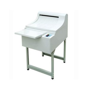 Model PLX-380H Film Format X-ray, CT, MRI and other medical films Film Size 3″ x 6″ ~ 14″ x 17″ Maximum Processing Width 14′ Adjustable Processing Time 25s, 35s, 45s Processing Speed 90s ~ 165s (optional) Drying Temperature Adjustment 40~60°C Liquid Chemical Cooling Temperature Adjustment 20~40°C Tank Capacity 6L Replenishment System Automatic Power Supply AC220V, 50Hz/10A single-phase Noise Level ≤55db Maximum Processing Capacity 160pcs (14’x17′), 180pcs (10’x12′)
Automatic X-ray Film Processor
-
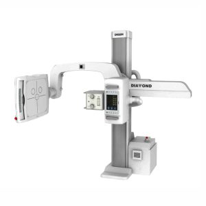 DrGem Diamond All-In-One Digital X-ray Machine is a fully automatic digital radiography system providing state-of-the-art image quality, image processing and user interface. With a wide selection of anatomical studies on the imaging software, DIAMOND automatically sets up the x-ray generator’s pre-programmed exposure technique settings, motorized radiographic stand positioning, x-ray collimation and post-image processing for the selected study. Specifically designed to increase workflow, this fully digital system offers convenient auto-positioning and advanced image processing to achieve big performance with little effort. Features of DrGem Diamond All-In-One Digital X-ray Machine: Outstanding Image Quality – Digital radiography via at panel detector improves your workflow, exam speed and comfort with efficiency. Digital at panel detector with Csl screen provides excellent spatial resolution, MTF, DQE and stability based on ne pixel pitch. A 3-field ion-chamber is provided for AEC function. Automatic Collimation – Automatic x-ray eld size control of the motorized collimator corresponds to dierent SIDs. Includes user adjustable lamp timer with on/oswitch. Automatic Positioning – DIAMOND is a fully-automatic diagnostic system with motorized movement and pre-programmed data for automatic positioning that can be easily reprogrammed by the operator. Seven safety sensors have been integrated into the DIAMOND system to protect patients from accidental collision. Automatic Stitching – DIAMOND 5A provides outstanding automatic stitching function via source tilting for creating one long composite image. DIAMOND Positioning Guide – The U-arm rotation ranges from +120 ° to +30 ° with SID movements from 100cm to 180cm. The mobile patient table can also be used to take patient images in a variety of positions for a total of over fifteen different positions (Chest PA, Skull Towne’s, Abdomen Supine, Hand AP, C-T Spine Swimmers and Abdomen Decubitus etc.) Included – Software, HP Laptop Computer – CPU≥3.2GHz – Memory capacity:≥4GB – Hard drive capacity :≥500 GB – Resolution: 1280 x 1024 – Display size: 21 inch color LCD screen – 64 bit Windows 10 operation system – Core: i5 Technical Specification: Power Rating – 52KW mA – 10 to 640mA mAs – 0.1 to 500mAs kV – 40 to 150kV, 1kV Step Generator – GXR-52 Rotor – Dual Speed (High and Low) Input Power – 400/480VAC±10%, 3Ø Line Frequency – 50/60Hz X-ray tube – DXT-12M 0.6/1.2mm, 300kHU Click Here To Download Catalogue
DrGem Diamond All-In-One Digital X-ray Machine
-
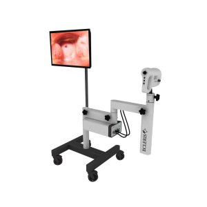 The Ecleris ColpoHD Digital Video Colposcope offers clinicians an illuminated, high definition magnified view of the cervix and tissues of the vagina and vulva. As a screening tool for sexual assault victims or for the early detection of precancerous lesions, the ColpoHD provides insight into relevant pathology changes of tissue shape and color with magnification of up to 50x and a digital zoom up to 128x. The integrated electronic green filter delivers exceptional pathology visualization, with clear, true color imaging and superb depth of field. The swing arm mounted high-definition (HD) camera can be easily maneuvered into place. Optional integrated image capture is available for documentation along with archiving and printing of images. A powerful tool for cervical cancer screening The ColpoHD is precise and easy to handle in all examination situations. High quality optics along bright LED illumination and high-tech CMOS image sensors guarantee fantastic quality HD images are displayed for every patient. The ColpoHD is also ideally suited for the examination of sexual assault victims who may be uncomfortable with a traditional Colposcope examination with the physician working through the standard microscope binoculars. The ColpoHD offers distance and a level of privacy for these patients who may be in a vulnerable frame of mind. The ColpoHD also offers optional image archiving and documentation of the examination in high definition image quality which is vital for sexual assault cases. Features at a Glance Outstanding image quality. Perfect HD 1280X720p color definition. Optical zoom. LED high power illumination. Great maneuverability. OSD (on screen display) with indication of real magnification. Zoom and focus operated by 4 commands joystick. Still function. WB (white balance) push bottom control. Perfect color discrimination due to CMOS HD technology. Floor stand system with 4 antistatic wheels with break. Ratched up down pole with double arm for easier and steady positioning. Magnification progressive. Optical zoom up to 37x and digital zoom up to 70x. Focus automatic and manual. Distance 240-300mm. Filter optical green. Led life 50.000 hs. Optional: monitor stand pole / Endodigi HD recording device. Technical Specification Outputs – 2x HDMI Magnification – Progressive magnification. Optical zoom up to 50x and digital zoom up to 128x. Focus – Automatic and manual Resolution – 1280 x 720 P Depth of Field – 250 – 300 Field of View – 8 – 140 mm Distance – 200 – 320 mm Filter – Optical green Light Source – LED High Intensity, 6500 Kelvin LED Life – 50.000 hrs Illuminated Control – Electronic adjustment. Constant light color. Power Supply – 100 – 120 VAC, 50/60 Hz Content Includes: Floor Mounted with Base with 4 Castors, Video Head, Positioning Arm, LED Light Included on Video Head, 110/220V Power Cable and User´s Guide, Stand For LCD Monitor and Printer, Digital Capturing System for Images, Videos and Sounds, USB 2.0. Includes Main Unit Processor with three Camera Inputs, USB Cable, Software, Hands Free Microphone and Footswitch. Click Here To Download Catalogue
Ecleris HD Digital Video Colposcope
-
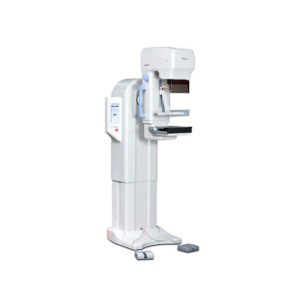 MX-600 is designed compactly for easy install and operation. Features: Auto Standard Positioning system (ASP) With Auto Standard exposure Positioning systems designed to maximize the convenience of radiography, you can easily adjust positioning by using standard exposure (RCC, LCC, RMLO, LMLO). ASP function makes operators easy to execute 4 axis exposures by software programming. One-touch button controls 4 standard positions (RCC, LCC, RMLO, LMLO) using C-Arm pre-set adjustment (MedioLaterral, Oblique view and CranioCaudal view). The ISO level can adjust the level of MX-600 standing position when it operates from vertical exposure to oblique and vice versa. Intelligent Automatic Exposure Control (AEC) Automatic Exposure Control system enables production of images with reliable intensity for any film, screen, or digital radiography. Furthermore, it greatly enhances the convenience of radiography by embedding the Full-AEC function which utilizes Auto kV control. It sets the best diagnosis environment by reducing manual operation and increasing patient throughput. Compression with comfort in mind When pressure is required for Mammography, it allows you to apply appropriate amount of pressure (up to maximum of 20kg) and is equipped with MICOM control’s Soft-touch system which minimize the discomfort. Automatic Conversion (Filter) Motorized and manual breast compressions are available for MX-600. MX-600 displays compression level and thickness on main body’s panel. Stable Output A molybdenum (0.03mm Mo) and Aluminum (0.5mm Al) filter are installed to absorb unnecessary x-ray. Mo filter covers low level kV range (22-35kV) and Al filter covers high kV range (35-39kV). Mo filter is useful for increasing image contrast in large breast with large amounts of glandular tissue. The rhodium filter (0.025mm Rh) can be installed as an option (as replacement of Al filter). Display of Exposure Condition When some degree of pressure is required for radiography, it allows you to apply the appropriate pressure (up to a maximum of 20kg) and is equipped with MICOM control’s Soft-touch system which is designed to minimize the discomfort of the examine with in the pressure range. Technical Specifications: Generator Type: High frequency inverter (40 kHz). Radiographic qualification: Great focus 22-39kV / 1-600mAs. Small bulb 22-35kV / 1-100mAs. Tube Focal point size: 0.1 / 0.3mm. Anode heat capacity: 300KHU (molybdenum). Filtration: Mo. Radiographic support Movement up / down: 720-1420mm (motorized). Rotating movement: ± 180˚ (Motorized) . Automatic standard positioning (ASP). SID: 650 mm. Bucky device Cassette size: 18x24cm, Grid: 4: 1, 91 lines / inches. Automatic exposure control (AEC) Type: solid state detector. Mode: 3 modes (Full / Semi / Manual). Density adjustment: 19 steps. Collimator: Automatic. Accessories Face protection. Film marker, hand switch, point compression paddle. 18x24cm Bucky device with compression paddle. Collimating device (18×24 and dotted plate). Options Set of Bucky devices of 24×30 cm. Set of 1.5X or 1.8X magnification devices. Click Here To Download Catalogue
Genoray Analogue Mammography MX-600
-
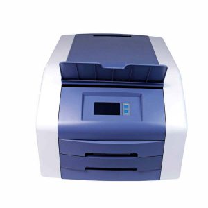
 The HQ-760DY Dry Imager is a thermo-graphic film processor designed for digital radiography imaging. It is the one and only domestically engineered medical dry thermal imager. The HQ-DY series Dry Imager uses the latest direct dry thermal imaging technology which accommodates for a full range of applications, including CT, MR, DSA and US, as well as CR/DR applications for GenRad, Mammography, Orthopaedics, Dental Imaging and more. The HQ-Series Dry Imager dedicates to precision in diagnosis with its outstanding image quality, and offers affordable imaging catering to your needs. – Supports Mammography – Dry thermal technology – Daylight load film cartridges – Double tray, supports 4 film sizes – Speed printing, higher efficiency – Economical, stable, reliable – Compact design, easy installation – Straight forward operation, user-friendly Technical Specification: Print Technology Direct thermal (dry, daylight-load film) Spatial Resolution 508dpi (20pixels/mm) Grayscale Contrast Resolution 14 bits Film Tray Two supply trays, 200-sheet capacity total Film Sizes 8’’×10’’, 10’’×12’’, 11’’×14’’, 14’’×17’’ Applicable film Medical Dry Thermal Film (blue or clear base) Interface 10/100/1000 Base-T Ethernet (RJ-45) Network Protocol Standard DICOM 3.0 connection Image Quality Automatic calibration using built-in densitometer Control Panel Touch Screen, Online Display, Alert, Fault and Active Power Supply 100-240VAC 50/60Hz 600W Weight 50Kg Operating Temperature 5℃-35℃ Storage Humidity 30%-95% Storage Temperature -22℃-50℃ Click Here To Download Catalogue
HU.Q hq-760dy Dry Image X-Ray Film Printer (Mammo)
-
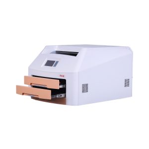
 Signers Digital Dry Thermal X-ray Film Printer – Is a dry imager designed to output information processed through DICOM network protocol, supports 4 films sizes. 8”x10”, 10”x12”, 11”x14” and 14”x17” Achieve the convenience of multiple sizes. Technical Specifications: Print Technology – Direct thermal (dry, daylight-load film) Spatial Resolution – 320dpi (12.6 pixels/mm) Throughput – 14”x17” ≥ 50 sheets/h, 8”x10” ≥ 70 sheets/h Film Tray – 2 supply tray Gray scale Contrast Resolution – 14 bits Network Protocol – Standard DICOM 0 connection Film Transferring Method – Rubbing Click Here To Download Catalogue
SIGNERS SUPiA Dry Thermal X-ray Film Printer
-
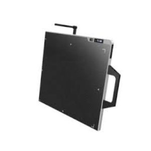 Features: Convenience: * AED (Automatic Exposure Detection) * Up to 3 detectors can be connected * Emergency Study * Optimized digital retrofit solution for X-ray system * Led lamp applied Speed: * Cycle time: less than 4.5 sec. * 1Gbps Ethernet * Steller gram, our console software, is simple, fast, stable and compatible * Promix: Signers processing tool * Client viewer (up to 5 places connectable, coming soon) Standard Accessories: Stellar M17 1Pc. DR: 17″x17″ Wired Detector CSI with Stellar Gram S/W 1Pc. Specification: Sensor: Amorphous Silicon Scintillator: CSI (Cesium-Iodide) Active Pixel Area: 427 x 427mm Pixel Pitch:139µm Resolution: 3.6 Ip/mm Pixel Matrix: 3072 x 3072 A/D Conversion:16 bit Cycle Time: <4.5 sec Interface (Wire):1Gbps Ethernet Dimension: 460mm(W) x 460mm(L) x 15mm(H) Weight: Detector: 4.0kg Console box: 1.1kg Operation Environment:50C to 350C, 30% to 85% RH Power: DC 24Vdc, 1.25A PC Min. Requirements: CUP: more than Intel Premium G5400 RAM: more than 8GB OS: Win 10 pro, 64bit Ethernet Card: Intel Gigabit Monitor: more than 17” Click Here To Download Catalogue
Stellar M17 CSI 17”x17” Wired Digital Radiography
-
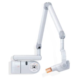 The ESX s tandard x-ray is an intraoral x-ray imaging system which offers safety, reliability, and greater functionality. It also features a multidirectional full- swivel revolution. The tube head revolves freely around the sphere, so practically any position can be achieved, including complete vertical m ovement. What is the ESX? DC type high frequency intraoral x-ray system Intuitive & easy to use operating panel Smart & reliable de sign for easy & steady use Automatic & optimal capture setting Support b oth of film and digital sensors Flexible i nstallation ( chair/ Wall type)
Vatech Standard X-ray System ESX Series
-
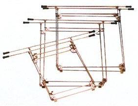 Lead Apron Lead Gloves Thyroid Shield Gonad Shield (Set of Three) Lead Protection Screen 3 x 6 Feet Lead Protection Screen 3 x 6 Feet Fim Markers Complete Set Safe Light Lead Glass 80 x 100 cm (8MM) Lead Glass 35 x 35 cm (8MM) Lead Glass 22 x 22 cm (8MM) Lead Sheet 4 X 6 Feet Each Viewing Boxes Single Each Viewing Boxes Double Each
x-ray hangers
Zenrox Healthcare Solutions - Advanced Medical Equipment in Lagos
Zenrox offers cutting-edge medical equipment and comprehensive support services in Lagos. Explore our products designed for superior healthcare delivery. Contact us today for innovative medical solutions.